Case Report, Res Rep Gastroenterol Vol: 2 Issue: 1
Perforated Sigmoid Volvulus in an Elderly Male with Expressive Aphasia
Steven KM Vuu, Tania Zavalza Jimenez, Carlos E Ramirez, Michael J DeRogatis* and Gudata Hinika
Department of Surgery, Dignity Health California Hospital Medical Center, Los Angeles, CA, USA
*Corresponding Author : Michael J DeRogatis
Department of Surgery, Dignity Health California Hospital Medical Center, Los Angeles, CA, USA
Tel: +1 4849035655
E-mail: michael.derogatis@gmail.com
Received: June 27, 2018 Accepted: July 10, 2018 Published: July 16, 2018
Citation: Vuu SKM, Jimenez TZ, Ramirez CE, DeRogatis MJ, Hinika G (2018) Perforated Sigmoid Volvulus in an Elderly Male with Expressive Aphasia. Res Rep Gastroenterol 2:1
Abstract
We report on a 73-year-old male with expressive aphasia and multiple comorbidities that presented with generalized abdominal pain, nausea, and no bowel movement or flatus in the last 3 days. On physical exam his vital signs were stable and his abdomen was distended, firm, and diffusely tender to palpation with guarding along with hyperactive bowel sounds. Computed Tomography (CT) scan was highly suggestive of a sigmoid volvulus. Colonoscopic decompression was unsuccessful, so surgical repair of the volvulus was indicated. Laparotomy revealed significant distention of the sigmoid and rectosigmoid junctions with a sigmoid volvulus that was impacted with stool up to the ascending colon, and a small serosal tear to the cecum with bowel contents dispersed throughout the abdomen. Rectosigmoid resection with a diverting colostomy and an ileo-ascending colon anastomosis were performed. It is important to recognize the constellation of symptoms for sigmoid volvulus in patients of this population in order to reduce incidence and progressive consequences associated with chronic presentation.
Keywords: Sigmoid volvulus; Computed tomography; Laparotomy
Introduction
A volvulus is in reference to the torsion of a segment of the alimentary tract on the axis formed by the mesentery. The most common sites of volvulus are the sigmoid colon (60.9%) and the cecum (34.5%), but can also occur in the transverse colon (3.6%) and splenic flexure (1%) [1,2]. First described by Rokitansky in 1836, a sigmoid volvulus occurs when an air-filled loop twists around the mesentery [3]. Sigmoid volvulus is the second most common cause of complete colonic obstruction and classically described as an illness of elderly, immobile, and persons with psychiatric disorders. Generally symptoms of abdominal pain, constipation, and nausea are reported. Other causes of colonic obstructions are adenocarcinoma and scarring secondary to diverticulitis. It is important to note patient age and comorbidities when considering differential diagnoses and patient workup.
Case Report
A 73 year old male with a past medical history of multiple cerebrovascular accidents with expressive aphasia, left arm contracture, end stage renal disease on hemodialysis, diabetes mellitus and past surgical history of laparoscopic appendectomy in 2016 and right inguinal hernia repair with mesh in 2015 stated that 5 days before admission, he had nausea and intermittent generalized abdominal pain. His last bowel movement was 3 days prior to admission and his vitals were stable. On physical examination, bowel sounds were hyperactive, the abdomen was distended, firm, and diffusely tender to palpation with guarding. The labs were all within normal limits without leukocytosis. The patient was subsequently taken for a computed tomography (CT) of the abdomen without contrast due to his renal failure, which revealed an extremely distended right colon (Figures 1-3). The caliber of the lumen was 12.5 cm and on abdominal x-ray, the colon had a coffee bean appearance (Figure 4). Impacted fecal material in the distended sigmoid and distal colon were also noted (Figures 1 and 2). Transverse view showed multiple air fluid levels indicative of a bowel obstruction (Figure 3). This imaging was concerning for a sigmoid volvulus. An unsuccessful colonoscopic decompression was attempted which revealed severe fecal impaction. Surgical repair of the volvulus was implicated and the patient was placed on strict order for nothing by mouth and rehydrated with intravenous fluids in anticipation for surgery. The risks, benefits, complications, and alternatives were discussed with the patient and his family. The patient consented to undergo exploratory laparotomy with bowel resection and a diverting colostomy.
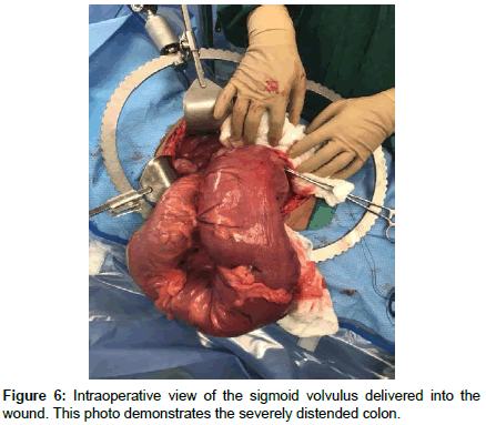
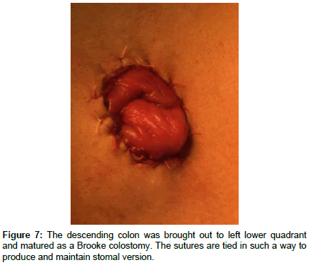
He was brought to the operating suite where he was positioned, prepped, and draped in a sterile fashion and intubated under general anesthesia. A midline incision was made into the peritoneal cavity. There was significant distention of the sigmoid and rectosigmoid junctions with a sigmoid volvulus going all the way to the right upper quadrant and covering most of intra-abdominal content. A colostomy to evacuate air was not successful because the volvulus was not filled with air but rather cemented with stool (Figure 5). At this point, the sigmoid volvulus was delivered into the wound (Figure 6). The descending colon was resected proximally and the upper rectum distally using a GIA stapler. The mesentery of the sigmoid colon and descending colon was taken down using a ligature device. There was also a small serosal tear to the cecum, so an ileo-ascending colon anastomosis was done using the GIA stapler. Copious irrigation of the intra-abdominal content and hemostasis was achieved using Surgicel and electrocautery. The descending colon was brought out to left lower quadrant and matured as a Brooke colostomy (Figure 7). We changed our gowns and redraped the abdomen and then turned to closing the midline fascia defect with 0 PDS in a running fashion. We used a retention suture of all vicryl in an interrupted fashion. Again, we copiously irrigated the wound. A Jackson-Pratt (JP) drain was placed in the abdomen to the right lower quadrant. The patient tolerated the procedure well and was monitored in the intensive care unit (ICU) due to his multiple comorbidities.
On postoperative day 1, the patient was doing well and was continuing with nothing by mouth, intravenous antibiotics, and drains. By postoperative day 2, the patient was out of the ICU and was transferred to the med-surg floor. By postoperative day 3, the patient was producing bowel movements into the colostomy bag. He was discharged on postoperative day 7 to go home with nursing care. At his 6 month follow up, he was doing well with no new complications.
Discussion
Sigmoid volvulus occurs when an air-filled loop twists around the mesentery. Interestingly, our patient had a sigmoid volvulus that was comprised of stool. Chronic constipation may cause elongation and dilatation of the sigmoid colon, therefore predisposing the patient to this condition. Colonic dysmotility is also a risk factor for sigmoid volvulus because it can cause chronic constipation. As in our patient with multiple comorbidities, he had a severe fecal impaction due to dysmotility. It is important to note that even if the patient has passed stool, the possibility of constipation may still exist.
Obstruction of the intestinal lumen occurs when the degree of torsion exceeds 180 degrees and decreased vascular perfusion occurs when the torsion exceeds 360 degrees. In the United States, sigmoid volvulus accounts for less than 10% of cases of intestinal obstruction, but accounts for 50%-80% of cases of intestinal obstruction in other places of the world. Although it can occur in younger patients and children who have abnormal colonic motility, sigmoid volvulus is mostly seen in older adults who are debilitated due to psychiatric or neurological conditions [1]. Other risk factors include previous episodes of volvulus, previous abdominal operations, and megacolon most often from Chagas disease or Hirschsprung disease.
Patients with sigmoid volvulus typically present with slowly progressive and constant abdominal pain, nausea, abdominal distension, and constipation, as our patient did. An abdominal computed tomography (CT) scan is used to make the diagnosis of sigmoid volvulus. A whirl pattern that is caused by the dilated sigmoid colon, mesocolon, and it’s vessels is noted, along with a bird beak appearance at the proximal and distal ends of the sigmoid volvulus [4]. In 60%-75% of cases, abdominal x-rays can be diagnostic with classic findings such as the “coffee bean sign” [1]. If there is also an incompetent ileocecal valve, one can also find a distended small bowel.
Flexible sigmoidoscopy can be used to reduce a sigmoid volvulus, but surgery may be required to prevent recurrent volvulus [1]. Recurrence in those who have not had surgery is estimated to be up to 90% [1,5]. Immediate laparotomy is performed when there are signs of peritonitis or if endoscopic detorsion is unsuccessful. Endoscopic detorsion using an endoscope is reported to be successful in 70%- 80% of the patients [1]. A sigmoidoscope can be used to detorse the sigmoid volvulus and can be further advanced into the twisted colon, restoring blood supply. In our patient, endoscopic detorsion was unsuccessful due to the severity of fecal impaction.
Emergent Surgery is indicated if there are signs of sepsis, gangrenous bowel is noted, or peritonitis. Studies show that emergent surgery has a mortality rate of 6.6 % and it raises to 11 % if gangrenous bowel is present [1,5]. Hartmann’s procedure, where the proximal rectal stump is closed and the distal colon is matured as a colostomy, should be performed if there are any factors that can threaten the viability of an anastomosis [1]. Although, studies show that emergent surgery has a mortality rate of 6.6% and it raises to 11% if gangrenous bowel is present [5].
Conclusion
It can be difficult to diagnose a sigmoid volvulus in elderly patients with expressive aphasia. Abdominal symptoms must be taken seriously and careful observation of bowel habits from the caretakers can help prevent a patient of being in grave danger from the consequences of a perforated bowel.
References
- Gingold D, Zuri M (2012) Management of colonic volvulus. Clin Colon Rectal Surg 25: 236-244.
- Lawrence PF, Bell RM, Dayton MT (2013) Colonic obstruction and volvulus. Essentials of General Surgery (5th edtn), Lippincott Williams & Wilkins, Philadelphia, USA.
- Atamanalp SS (2010) Sigmoid volvulus. Eurasian J Med 42: 142-147.
- Da Fonseca LM, Caldeira DA (2013) The whirl CT sign in patient with sigmoid volvulus due chagas’ disease. Indian J Surg 75: 162-163.
- Williams M, Steffes CP (2006) Sigmoid volvulus in a 46 year old man. Case report and literature review. J Hosp Phys 1: 33-36.
 Spanish
  Chinese
  Russian
  German
  French
  Japanese
  Portuguese
  Hindi
Hindi 


