Case Report, Int J Ophthalmic Pathol Vol: 12 Issue: 4
Pebble in the Well-Myopia Due to Inflammation of Retina and Choroid-Unique: Case Report
Aliya Sultana*
Department of Ophthalmology, Osmania University, Telangana, India
*Corresponding Author: Aliya Sultana
Department of Ophthalmology, Osmania
University, Telangana, India
Tel: 7337419409;
E-mail: draliyasultana23@gmail.com
Received date: 29 January, 2023, Manuscript No. IOPJ-23-88167;
Editor assigned date: 31 January, 2023, PreQC No. IOPJ-23-88167 (PQ);
Reviewed date: 14 February, 2023, QC No. IOPJ-23-88167;
Revised date: 20 April, 2023, Manuscript No. IOPJ-23-88167 (R);
Published date: 27 April, 2023, DOI: 10.4172/2324-8599. 12.4.030
Citation: Sultana A (2023) Pebble in the Well-Myopia Due to Inflammation of Retina and Choroid-Unique: Case Report. Int J Ophthalmic Pathol 12:4.
Abstract
60-Years-old male patient presented with diminished vision in right eye since many years, left eye since one year. He is security guard by occupation. No history of trauma. Not using spectacles for distance vision. On examination right eye BCVA is no perception of light, deep anterior chamber, mid dilated, fixed pupil, aphakia, and fundus was mild vitritis, dislocated crystalline cataractous lens which was wandering or floating in the vitreous cavity with the position, pale disc, pigmentary changes in posterior pole and peripheral retina. Left eye BCVA is 1/60, anterior segment is normal, pupil was reacting to light immature senile cataract, fundus was media hazy due to cataract, disc was normal with chorio retinal degenerative changes in posterior pole and peripheral retina. Right eye has no prognosis so management not done where as in left eye cataract surgery done, post-operative vision after six weeks was 6/18, patient was advised to come for regular follow-ups to detect early if any macular changes progress to macular exudation and neovascularisation like choroidal neo vascular membrane due to age related macular degeneration or myopia.
Keywords: Myopia, Inflammation, Uveitis, Zonules, Sub Retinal Fibrous Bands (SRFB), Dislocated lens
Introduction
When the growth of the eyeballs is in the same amount then symmetric myopia occurs, if the axial growth or the combined refractive power of the cornea and lens is different between two eyes will cause asymmetric myopia or anisometropic myopia [1]. Axial and refractive myopia can be differentiated by measuring corneal power and axial length. In these cases, there will be monocular visual deprivation due to high myopia in one eye. Uniocular high myopia is usually associated with many congenital anomalies like optic nerve, lens and macular abnormalities, in our case dislocated lens and macular scar noted [2]. Sometimes extensive axial length may be due to abnormal central brain mechanism which controls the ocular growth. Various environmental factors and genetic factors are involved in progression of myopia [3].
Myopia increases the risk of pathological ocular changes such as cataract, glaucoma, and retinal detachment, which can cause irreversible vision loss [4].
Case Presentation
60-years-old male, security guard by profession, presented with diminished vision in both eyes, Right Eye (RE) since 15 years and in Left Eye (LE) since one year, family history was not significant, no history of trauma, birth history was normal and no history of using spectacles previously for distance vision [5]. Systemic examination was normal. Patient underwent detailed ocular examination and documentation [6].
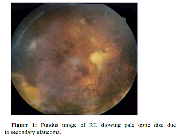
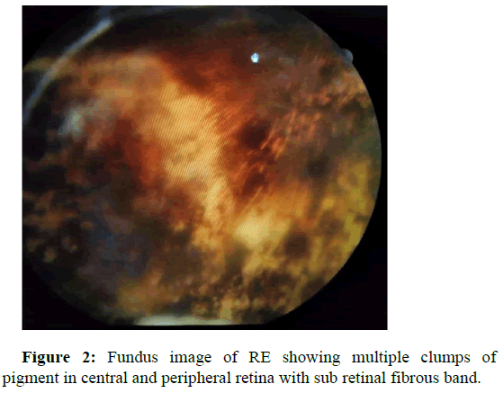
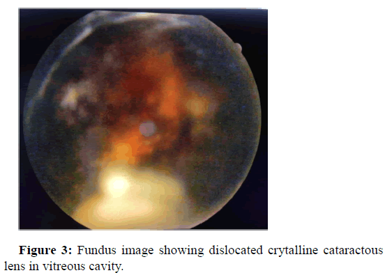
BCVA in RE was no perception of light, slit lamp examination showed deep anterior chamber with fixed, dilated pupil, aphakia and fundus revealed hazy media due to vitritis, pale disc, disc gliosis, multiple pigmentary clumps of various sizes in central and peripheral retina, sub retinal fibrous bands and floating or wandering crystalline cataractous lens in the vitreous cavity in relation to the position of the patient (Figures 1-3).
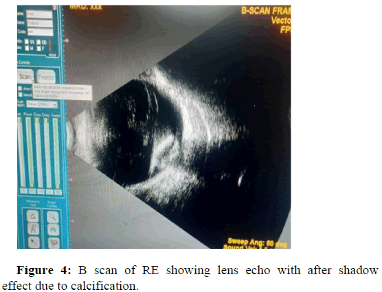
B scan of RE showed lens echo with after shadow effect, probably due to calcification of lens (Figure 4).
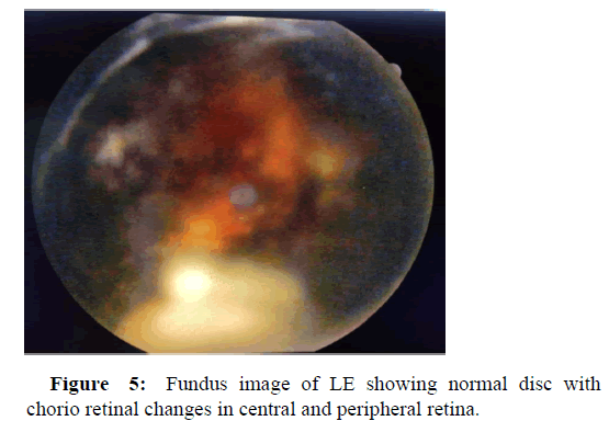
Left eye BCVA was 1/60, anterior segment was normal, lens was cataractous, fundus was hazy due to lenticular opacity, and disc was normal, chorio retinal changes in the central and peripheral retina, sub retinal fibrous bands in the periphery (Figure 5). LE cataract surgery done, post-operative BCVA after 6 weeks was 6/18.
Discussion
Myopia is a global health public problem, which not only progresses during young age, also can be progress in old age. Myopia is bilateral refractive error, sometimes one eye has minimal refractive error and the other eye will be highly myopic. Today we have various methods to control myopia progression and prevent the blindness. In our case I want to bring one point into consideration that inflammation has very important role in progression of myopia, inflammatory cytokines are the mediators which are released due to retinal inflammation like retinitis or chorio retinitis can lead myopia progression, we noticed sequalae of chorio retinitis like sub retinal fibrous bands and pigmentary chorio retinal changes which indicates the healed or resolved chorio retinitis. More changes in RE, may be due to severe chorio retinitis in RE compared to LE.
Myopia increases the risk of pathological ocular changes such as cataract, glaucoma, and retinal detachment, all of which can cause irreversible vision loss.
Most inflammatory conditions related to myopia involve the chorio capillaries. Clinical conditions like multifocal choroiditis and/or punctate inner choroiditis, multiple evanescent white dot syndrome, acute idiopathic blind spot enlargement and VKH. Anatomical changes due to inflammation induced myopia will cause fragility of the chorio capillaries and lead to inflammatory Choroidal Neo Vascular Membrane (CNVM).
Possible mechanism for the myopic shift is supraciliary exudation which causes relaxation of zonular fibers and increased convexity of the crystalline lens.
Corticosteroid lens changes is accompanied by a marked increase in lens induced myopia up to more than 7 diopters due to the optical effect of the posterior sub capsular cataract. In our case patient did not give any history of treatment so drug induced myopic shift is not possible.
In axial myopia, ciliary zonules are poorly developed, myopic lenses weigh 5.5% less than normal lenses, which results in sub normal tension, dislocation of the lens in our case is probably due to sub normal tension by zonular dysplasia. In RE dislocated crystalline lens might have caused secondary glaucoma due to lens induced inflammation in vitreous cavity.
Conclusion
Highly myopic eyes have risk of many structural changes in the eye. Our case is diagnostic challenge case because we don’t know whether it is inflammation (uveitis) induced myopia or myopia induced inflammation. Structural globe changes due to myopia cause chorio capillaris/retinal pigmentary epithelium complex fragility, rendering it prone to inflammatory diseases. Patient presented to us after long time with many changes in retina, so it is confusing, the axial length of the globe in LE is normal with many chorio retinal changes, more in favour of inflammation induced chorio retinal changes, and in RE the axial length is slightly more compared to LE, possibly anisometropic myopia associated with lens and retinal changes. Both eyes are showing sub retinal bands in periphery with healed pigmented chorio retinal patches in all quadrants so we are analysing to be uveitis induced myopia.
References
- Weiss AH (2003) Unilateral high myopia: Optical components, associated factors, and visual outcomes. Br J Ophthalmol 87:1025-1031.
[Crossref] [Google Scholar] [PubMed]
- Shinojima A, Negishi K, Tsubota K, Kurihara T (2022) Multiple factors causing myopia and the possible treatments: A mini review. Front Public Health 10.
[Crossref] [Google Scholar] [PubMed]
- Herbort CP, Papadia M, Neri P (2011) Myopia and inflammation. J Ophthalmic Vis Res 6:270-283.
[Crossref] [Google Scholar] [PubMed]
- Gross SA, von Noorden GK, Jones DB (1993) Necrotizing scleritis and transient myopia following strabismus surgery. Ophthalmic Surg 24:839-841.
[Google Scholar] [PubMed]
- Koch HR, Siedek M (1997) Lens induced myopia in steroid cataracts (author's transl). Klin Monbl Augenheilkd 171:620-622.
[Google Scholar] [PubMed]
- McKanna JA, Casagrande VA (1978) Reduced lens development in lid-suture myopia. Exp Eye Res 26:715-723.
[Crossref] [Google Scholar] [PubMed]
 Spanish
  Chinese
  Russian
  German
  French
  Japanese
  Portuguese
  Hindi
Hindi 