Research Article, J Mar Biol Oceanogr Vol: 7 Issue: 2
Kinorhyncha Distribution in Intertidal Regions of South Andaman Regions
C. Jeeva, P.M. Mohan* and M. Muruganantham
Department of Ocean Studies and Marine Biology, Pondicherry University, Brookshabad Campus, Port Blair-744 112, Andaman and Nicobar Islands, India
*Corresponding Author : P.M. Mohan
Department of Ocean Studies and Marine Biology, Pondicherry University, Brookshabad Campus, Port Blair-744112, Andaman and Nicobar Islands, India
Tel: +91-3192-262317
Fax: +91-3192-262323
E-mail: pmmtu@yahoo.com; pmmnpu@rediffmail.com
Received: April 02, 2018 Accepted: May 14, 2018 Published: May 21, 2018
Citation: Jeeva C, Mohan PM, Muruganantham M (2018) Kinorhyncha Distribution in Intertidal Regions of South Andaman Regions. J Mar Biol Oceanogr 7:2. doi: 10.4172/2324-8661.1000191
Abstract
Kinorhyncha Distribution in Intertidal Regions of South Andaman Regions
The lesser known taxa of Kinorhyncha are exclusively marine, holobenthic, free-living, meiofaunal species found in all marine habitats. However, the literature review suggested that the information on geographical distribution and taxonomic status of Kinorhyncha is yet to be revealed in larger scale, that also from the tropical seas. Although earlier work was carried out during the year 1969 in the Indian Ocean, the continuity of research was found to be negligible in the latter years. This research article presents a few new reports of the Phylum Kinorhyncha for the waters of the Andaman Islands. The present study was evolved five new reports and one redescription of Kinorhyncha from Andaman Islands, India.
Keywords: Kinorhyncha; Intertidal; Indian Ocean; Andaman islands; Tropical seas
Introduction
The holobenthic Kinorhyncha is exclusively marine, free-living, meiofaunal species within a size range of 0.13-1.04 mm body length [1]. Their distributions are global, and are found in all marine habitats [2,3]. The Kinorhyncha was first described in 1851 i.e. almost 165 years ago [4]. However, the knowledge of these animals is far from complete due to the fact that very small communities of researchers are interested presently on this phylum (currently less than ten people worldwide working on this group). The earlier works revealed that the systematic carried out by Reinhard [5,6] was modified by Zelinka [7] under the monograph “Monographie der Echinodera”. Moreover, this work provided first time information on all aspects of the morphology and biology of this taxon [7]. During 20th century studies of Higgins had been contributed a larger level of taxonomical studies on Kinorhyncha. Studies of Higgins and co-workers after 1960 described 60 new species, re-described 14 species, and established six new genera [1,8-27]. During the last decade, several new species were described by various researchers [28-41]. Recently so many efforts have been made to provide the comprehensive checklist of Kinorhyncha. Sørensen [42] listed 205 species of Kinorhyncha described from adult specimens, whereas Neuhaus [1] listed 191 species of Kinorhyncha described from adult specimens, and 50 species of juvenile. However, molecular phylogenetic studies by Yamasaki et al. [43] and Sørensen et al. [44] and description of new species from various geographical locations to support the need for a periodical revision of the Kinorhyncha classification. Indian scenario of Kinorhyncha is very meager and have only a few dozen of literature on this tropical shallow-water ecosystems. The meiobenthic invertebrate group Cateria styx species has been reported from the east coast of India by Rao and Ganapati [45]. Ganapati and Rao [46] identified species of kinorhyncha present in Waltaire coast. Cateria gerlachi sp., were identified by Higgins [14] reported from the east coast of India. Pycnophyes sp., were collected from the west coast of India by Dovgal et al., [47]. The specimen of Kinorhyncha family Pycnophyes sp., was collected from the west coast of India [47]. Although earlier work was carried out by Higgins [15] in the Indian Ocean, the continuity of research was found to be negligible. According to Venkataraman et al. [48] only 9 species were reported in India till 1979. After 38 years, works on Kinoryncha in India has taken up and evolved a first report Echinoderes setiger [49] from the Andaman Islands [50]. Overall, at present only 11 species that are recorded in Indian waters.
Based on an exhaustive bibliographical survey and revision of taxon names, it was found that the Phylum Kinorhyncha comprises of 256 Species belonging to 29 Genera and 13 Families. The Families are distributed under three Orders, such as Echinorhagata Sørensen et al. [44], Kentrorhagata Sørensen et al. [44] and Xenosomata Zelinka [51]. Among the 256 valid species, the last four years alone reported 67 new species under this group along with two new Orders, three Families and nine new Genera.
Materials and Methods
The sediment samples were collected during the year of 2015 and 2016 according to this Island weather pattern. These Islands have rainfall for the first week of May to last week of December in every year. This rain was influenced by SW monsoon during the period May to August as First Phase of Monsoon (SWM) and the period September to December influenced by NE monsoon considered as Second Phase of Monsoon (NEM). The period January to April considered a Non Rainy Season (NRS). The NRS samples were collected during the month of February 2015 and 2016. SWM samples were collected during June 2015 and 2016 and NEM samples were collected from October 2015 and 2016. The study area was identified in five locations around the intertidal regions of Port Blair, South Andaman Islands (Figure 1). The station name and coordinate are as follows: Wandoor (Lat.11°85’10.74”N–Long.092°3, 7’13.28”E) Dollygunj (Lat.11°38’14.05”N–Long.092°42’10.80”E) Shippighat (Lat.11°36’18.26”N–Long.092°42’04.54”E), Chidiyatappu (Lat.11°30’21.28”N–Long.092°42’09.32”E) and Carbyns Cove (Lat.11°38’20.25”N – Long.092°44’54.24”E) South Andaman Islands, India.
The quadrate method was adopted for the sediment sample collection. The plastic hand corer with a length of 15 cm and 6 cm diameter was used to collect the sediment samples from the study area. The corer was vertically buried into the sediment up to a depth of 5 cm and duplicate sediment samples were collected at each station. Each collected sediment samples was kept in polythene bag with 5% buffered formalin and 1% Rose Bengal.
The collected sediment samples were sieved through standard sieves (ASTME) to retain the 0.5 to 0.063 mm size ranged meiofauna with filtered seawater. The sieved meiofauna were identified under different group level under stereomicroscope (Nikon SMZ-1500), using the key of Higgins and Thiel [52] and Giere [53]. Among the observed meiofaunal community, the Kinorhyncha group as enumerated to the species level under a digital microscope (Carl Zeiss-Axiovert-40CFL).
Result
Echinoderes capitatus (Figures 2A-2D)
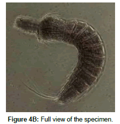
Diagnosis: This specimen’s head consists of a retractable mouth cone and oral stylets, but their exact number and arrangement could not be determined due to the retracted position of a captured animal. Head and Neck region are not considered as a segment. Total trunk length (TL) is 265 μm long and Mean Sternal Width (MSW-6) is 49 μm and which has MSW-6/TL percentage is about 18.49. The Standard Width (SW-10) is having – 44.47 μm which has 16.8% of trunk length (SW-10/TL). Trichoscalid spinoscalid plates and outer oral stylet are present, but it is not clearly visible. Midventral placid wider, 13.40 μm long and other placid are small and has 8.61 μm long. Trunk transverse section show triangular shape. Pectinate fringes moderately and visible to entire segment. Mid-terminal spine and mid-dorsal spine absent and first two trunk segment consist of a closed cuticular ring. Segment 2, 5, 8 and 10 exhibited latero ventral spines.
Description
S1 Segment 1 (S1) showed 13.49 μm long and closed cuticular ring visible and pachycyclus well developed. There are no middorsal spine, latero ventral spine and sensory spots.
S2 Segment 2 (S2) showed a length of 12.37 μm and the ventro lateral tubules has a length of 12.19 μm long was observed. Pachycyclus well developed. The cuticular ring was closed ventral side of the specimen and not shows any middorsal spine and latero ventral spine.
S3 Segment 3 (S3) exhibited a length of 22.09 μm, with two sternal plate on ventral side and one tergal plate. Pachycyclus similar to preceding one, there is no middorsal spine and latero ventral spine.
S4 Segment 4, (S4) was having a length of 19.82 μm. Pachycyclus, sternal and tergal plate are similar to preceding one, there is no middorsal and latero ventral spine.
S5 Segment 5, (S5) had a total length of 28.96 μm and its latero ventral spine show a length of 16.01 μm, length. The pachycyclus, sternal and tergal plate are similar to preceding one and mid-dorsal spine absent.
S6, Segment 6 (S6) showed a length of 28.1 μm. The pachycyclus, sternal and tergal plate are similar to preceding one and one pair of glandular cell outlet present on dorsal side of the specimen. There were no middorsal spine and sensory spots.
S7 Segment 7 (S7) exhibited a length of 29.32 μm long. The pachycyclus, sternal and tergal plate similar to preceding segment and one pair of glandular cell outlet present on dorsal side of the specimen. There were no middorsal spine and sensory spots.
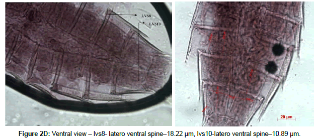
S8 Segment 8 (S8) had a length of 25.97 μm long with a Latero ventral spine 18.22 μm. The pachycyclus, sternal and tergal plate are similar to preceding one and one pair of glandular cell outlet present on dorsal side of the specimen. There are no middorsal spine and sensory spots.
S9 Segment 9 (S9) exhibited a 28.51 μm length. Pachycyclus, sternal and tergal plate are similar to preceding one and one pair of glandular cell outlet present on dorsal side of the specimen. There were no middorsal spine and sensory spots.
S10 Segment 10 (S10) had a length of 30.77 μm, long with a latero ventral spine 10.89 μm pachycyclus, sternal and tergal plate similar to preceding one, one pair of glandular cell outlet present on dorsal side of the specimen. Middorsal and sensory spots are absent.
S11 Segment 11 (S11) showed a length of 20.23 μm with a lateral terminal spine length (LTS) of 130 μm. The percentage with LTS/TL with a 49.06% of trunk length. One pair of lateral terminal accessory spine (LTAS) 17.74 μm long. Further, there were no Lateral spine and middorsal spine. Terminal sternal slightly pointed.
Based on the external morphology the studied specimen was identified as an adult female.
Classification
Phylum: Kinorhyncha [4]
Class: Cyclorhagida [44]
Order: Echinorhagata [44]
Family: Echinoderidae (Bütschli, 1876)
Genus: Echinoderes (Claparède, 1863)
Species: Echinoderes capitatus [7]
This specimen deposited in the Department of Ocean Studies and Marine Biology, Pondicherry University, Port Blair (DOSMB, PU, PB) depository. The Specimen Number is: DOSMB 06002
Habitat: The mangrove sediments of Wandoor, South Andaman. Midtidal environment sediment, that also only in the study period of First Phase of Monosoon, this species was identified and reported.
Distribution: This species was identified in the regions of South Coast of Central Java, Indonesia mangrove region, Singapore, Indonesia, Malay Peninsular, Malayan Archipelago [54]. The present study reported from Wandoor (Lat.11°35’10.74”N – Long.092°37’13.28”E) South Andaman Islands, India.
Discussion: The Echinoderes capitatus lack of Mid dorsal spines, enlarged anterior true trunk segment are characteristic identification structure of this species [18]. Lateral Spines only present in the Segment 2 and 8. Lateral seta observed on either side of segment 10 [19,20], This speceiment has to be considered as a holotypic beceause only one specimen was collected and preserved. Further, the works of Higgins [18] and Hyus and Coomans [55] confirm the non availability of mid dorsal spine for this species.
Echinoderes peterseni (Figures 3A-3E)
Diagnosis: This specimen’s head consists of the retractable mouth cone and oral stylets are present, but their exact number and arrangement could not be determined due to submergence in mounting media. Head and Neck region are not considered as a segment. Total Trunk Length (TL) is 286 μm and Median Sternal Width (MSW-6) is 50.31 μm. The percentage of MDW-6 with TL is 17.59% The Standard Width (SW-10) is 52.33 μm and the percentage with TL is 18.30%. The trichoscalid, spinoscalid plates and outer oral stylet are present, but it is not clearly visible. Midventral placid wider (10.34 μm) and other placids are small (6.12 μm). Trunk transverse section show triangular shape. Midterminal spine is absent. The first two trunk segment exhibited a closed cuticular ring. Segment 5-9 shown a latero ventral spines. The all segments are pectinate fringe poorly developed.
Description
S1 Segment 1 (S1) showed a length of 14.25 μm with closed cuticular ring visible and pachycyclus well developed. There is no middorsal spine and sensory spots on middorsal position. The subventral sensory spot clearly visible. Pectinate fringe poorly developed. Scare present in the midventral of anterior margin.
S2 Segment 2 (S2) showed a length of 13.43 μm with well developed pachycyclus. The cuticular ring was closed on ventral side of the specimen. Middorsal spine is not observed. Pectinate fringe poorly developed. One pair of ventrolateral tubules present.
S3 Segment 3 (S3) exhibited a length of 14.79 μm with two sternal plate in ventral side and one tergal plate in dorsal side. The pachycyclus is similar to preceding one. There are no middorsal spine and latero ventral tubules or spine observed. Pectinate fringe poorly developed.
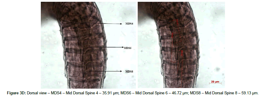
S4 Segment 4 (S4) was a length of 17.74 μm with the pachycyclus, sternal and tergal plates, which are similar to preceding one. Middorsal spine length is 35.93 μm and there is no latero ventral spine. Pectinate fringe poorly developed.
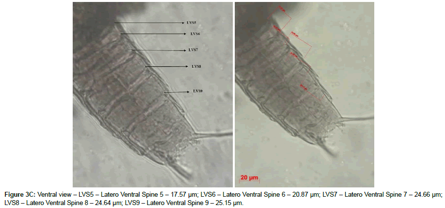
S5 Segment 5 (S5) had a length of 16.57 μm and its latero ventral spine show a length of 17.57 μm. The pachycyclus, sternal and tergal plate are similar to preceding segments and middorsal spine absent. One pair of cuticular scare presents on ventral side and one pair of subdorsal cuticular scare also present. Pectinate fringe poorly developed.
S6 Segment 6 (S6) showed a length of 18.49 μm with a latero ventral spine had a length of 20.87 μm. The pachycyclus, sternal and tergal plate are similar to preceding one and middorsal spine had a length of 46.77 μm. One pair of cuticular scar presents on ventral side and one pair of subdorsal cuticular scare were present. Pectinate fringe poorly developed.
S7 Segment 7 (S7) exhibited a length of 24.92 μm with 24.66 μm length of a latero ventral spine present. The pachycyclus, sternal and tergal plate similar to preceding one and middorsal spine was absent. One pair of cuticular scar presents on ventral side and one pair of subdorsal cuticular scare present. Pectinate fringe poorly developed.
S8 Segment 8 (S8) had a length of 23.21 μm with a length of 24.64 μm latero ventral spine present. The pachycyclus, sternal and tergal plate are similar to preceding one. The middorsal spine had a length of 59.13 μm. One pair of cuticular scar presents on ventral and subdorsal sides. Pectinate fringe poorly developed.
S9 Segment 9 (S9) exhibited a 26.39 μm length with a length of 25.15 μm of the latero ventral spine present. The pachycyclus, sternal and tergal plate were similar to preceding one. One pair of cuticular scar presents on the ventral as well as subdorsal sides and middorsal spine absent. Pectinate fringe poorly developed.
S10 Segment 10 (S10) had a length of 30.75 μm with the pachycyclus, sternal and tergal plate were similar to preceding one. The middorsal and lateral spines were absent. One pair of cuticular scar presents in ventral and sbudorsal sides. Pectinate fringe poorly developed.
S11 Segment 11 (S11) showed a length of 11.22 μm with a lateral terminal spine (LTS) length is 170.95. The percentage of LTS with TL was 59.77. One pair of lateral terminal accessory spine (LTAS) present with a length of 20.95 μm. Further, there are no lateral spine and middorsal spine. Terminal sternal slightly pointed. One pair of sensory spots presents on dorsal side of the specimen. Pectinate fringe poorly developed.
Based on the external morphology the studied specimen was identified as an adult female.
Classification
Phylum: Kinorhyncha [4]
Class: Cyclorhagida [44]
Order: Echinorhagata [44]
Family: Echinoderidae (Bütschli, 1876)
Genus: Echinoderes (Claparède, 1863)
Species: Echinoderes peterseni [56]
This specimen deposited in the Department of Ocean Studies and Marine Biology, Pondicherry University, Port Blair (DOSMB, PU, PB) depository. The Specimen Number is: DOSMB 06003
Habitat: The intertidal muddy sediments of Wandoor, South Andaman. Mid Tide environment represented this species. This specimen observed only in the study period of First Phase of Monosoon. Observed only one specimen in this study.
Distribution: This species was identified in Disko Island, West Greenland [56] and Arctic [57]. The present study reported from Wandoor (Lat.11°35’10.74”N– Long.092°37’13.28”E) South Andaman Islands, India. Only one specimen was collected.
Discussion: The Echinoderes peterseni mainly observed in the intertidal muddy region [56] which was matched with the present study also. Mid dorsal spine exhibited 4,6,8 segment in the species Echinoderes peterseni was also confirmed in the present studied species. This specimen also considered as a holotypic and preserved and deposited in the DOSMB, PU, PB. It is a really surprised observation that the Arctic species was observed in the tropical intertidal environment. At present the authors were not having any explanation for this observation. However, when looked in to the Andaman waters, a few reports such as a phytoplankaton species (Phaeocystis sp.) by Sachithanandam et al. [58], a Sabellariid Polychaete worm [59] and a Ascidian [60] from temperate region to tropical region was observed and recorded. So, this species was also considered a probality of temperate species occurrence in the tropical region. A further probe is needed for its occurrence or any morphological changes which was not identified by the present work.
Echinoderes horni (Figures 4A-4D)
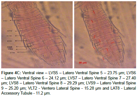
Figure 4C: Ventral view – LVS5 – Latero Ventral Spine 5 – 23.75 μm; LVS6 – Latero Ventral Spine 6 – 24.12 μm; LVS7 – Latero Ventral Spine 7 – 27.40 μm; LVS8 – Latero Ventral Spine 8 – 29.29 μm; LVS9 – Latero Ventral Spine 9 – 25.20 μm; VLT2 - Ventero Lateral Spine - 15.28 μm and LAT8 - Lateral Accessory Tubule - 11.2 μm.
Diagnosis: The head consists of the retractable mouth cone and oral stylets are present, but their exact number and arrangement could not be determined due to the submergence under the mounting media. Total Trunk Length (TL) was 257 μm and the Mean Sternal Width (MSW-6) was 45.86 μm. The percentage of MSW-6 with TL was 17.84. The Standard Width (SW-8) was 30.32 μm and its percentage with TL was 11.80. The Trichoscalid and spinoscalid plates, as well as outer oral stylet were present but not clearly visible. Midventral placid wider (8.72 μm) and other placid were small (5.91 μm). The trunk transverse section was in triangular shape. Pectinate fringe moderately visible to entire segment. Midterminal spine and middorsal spine absent and first two trunk segment were closed and represented as a cuticular ring. Segment 5-9 had latero ventral spines. The head and neck regions were not considered as a segment.
Description
S1 Segment 1 (S1) had a length of 14.80 μm and closed with cuticular ring. The pachycyclus was well developed. There were no middorsal spine and it had two sensory spots on middorsal position. The cuticular perforation producing a distinct pattern on each segment.
S2 Segment 2 (S2) exhibited a length of 14.89 μm and ventrolateral tubules has a length of 15.28 μm. The pachycyclus well developed and had a cuticular ring. There was no middorsal spine observed in this segment. The cuticular perforation producing a distinct pattern on each segment. Pectinate fringe moderately visible on posterior margins of the segments. Round cuticular spots present near anterior dorsal midline on segments 1-10.
The segments S3 to S10 represented the following characters over and above described in the individual species identification. They were the cuticular perforation producing a distinct pattern on each segment. Pectinate fringe moderately visible on posterior margins of the segments. Cuticular spots present on the anterio lateral on ventral surface of segments, nearer to the midline on segment 2-10.
S3 Segment 3 (S3) had a length of 20.34 μm. This segment had two sternal plate on ventral side and one tergal plate on the dorsal side. The pachycyclus similar to preceding one, there is no middorsal spine.
S4 Segment 4 (S4) had a length of 16.69 μm long, The, pachycyclus, sternal and tergal plate similar to preceding one, there is no middorsal and latero ventral spine.
S5 Segment 5 (S5) was having a length of 20.55 μm and the latero ventral spine had a length of 23.75 μm. The pachycyclus, sternal and tergal plate similar to preceding one, and middorsal spine absent.
S6 Segment 6 (S6) had a length of 15.68 μm with a latero ventral spine of length of 24.12 μm.The pachycyclus, sternal and tergal plates were similar to preceding one with middorsal spine absent.
S7 Segment 7 (S7) was a length of 20.14 μm with 27.4 μm long latero ventral spine. The pachycyclus, sternal and tergal plates were similar to preceding one, middorsal spine absent.
S8 Segment 8 (S8) with had a length of 20.80 μm, The latero ventral spine had a length of 29.29 μm. Lateral accessory tubule had a length of 11.2 μm and the long pachycyclus, sternal and tergal plates similar to preceding one, middorsal spine absent.
S9 Segment 9 (S9) had the length of 20.33 μm.The latero ventral spine exhibit a length of 25.20 μm. The pachycyclus, sternal and tergal plate are similar to preceding one and the middorsal spine absent.
S10 Segment 10 (S10) showed a length of 24.30 μm, and the pachycyclus, sternal and tergal plate similar to preceding one, middorsal and latero ventral spine absent.
S11 Segment 11 (S11) had a length of 12.22and , lateral terminal spine showed a length (LTS) 127.65 μm , and the percentage with TL was 49.68. Three pairs of penile spine called as (P1) 11.72 (P2) 16.09 and (P3) 9.42 μm are present. There were no lateral spine and middorsal spine. Terminal sternal slightly pointed.
Based on the external morphology the studied specimen was identified as an adult male.
Classification
Phylum: Kinorhyncha [4]
Class: Cyclorhagida [44]
Order: Echinorhagata [44]
Family: Echinoderidae (Bütschli, 1876)
Genus: Echinoderes (Claparède, 1863)
Species: Echinoderes horni [19]
This specimen deposited in the Department of Ocean Studies and Marine Biology, Pondicherry University, Port Blair (DOSMB, PU, PB) depository. The Specimen Number is: DOSMB 06004
Habitat: The intertidal sediments of Wandoor, South Andaman. Mid Tide environment represented this species. This speciemen observed only in the study period of First Phase of Monosoon. one speeimen was collected.
Distribution: This species was identified in the Twin Cays, Atlantic Barrier Reef [19], Florida [61], North East Atlantic [54] and Carrie Bow Cay [62], The present study reported from Wandoor (Lat.11°35’10.74”N – Long.092°37’13.28”E) South Andaman Islands, India. Only one specimen was collected.
Discussion: The Echinoderes horni observed in surface sediments of intertidal locations [19] was matched with the present study location. This species exhibit a smaller tergal extension [63] also supports the present identified species.
Echinoderes remanei (Figures 5A-5G)
Diagnosis: This specimen’s head consists of the retractable mouth cone and oral stylets are present, but their exact number and arrangement could not be determined due to its submergence in the mounting medium. Head and Neck region are not considered as a segment. Total Trunk Length is 260 μm long and Mean Sternal Width (MSW-6) is 52.15 μm. The percentage of MSW-6 with reference to TL is 20.05%. The Standard Width (SW-10) is 41.56 μm and the percentage with reference to TL is 15.98%. Trichoscalid, spinoscalid plates and outer oral stylet are present, but it is not clearly visible. Midventral placid wider (13.65 μm) and other placid are small (7.05 μm). Trunk transverse section show triangular shape. Pectinate fringes moderately and visible to entire segment. Mid-terminal spine absent and first two trunk segment consist of a closed cuticular ring. Segment 5-9 exhibited latero ventral spines.
Description:
S1 Segment 1 (S1) showed a length of 18.94 μm with a closed cuticular ring visible. The pachycyclus was well developed. There was no middorsal spine and it has two sensory spots on middorsal position.
S2 Segment 2 (S2) exhibit a length of 10.9 μm. The pachycyclus well developed. The cuticular ring was closed on ventral side of the specimen and not show any middorsal spine and latero ventral spine.
S3 Segment 3 (S3) exhibited a length of 16.97 μm, with two sternal plate on ventral side and one tergal plate on the dorsal side. The pachycyclus similar to preceding one, there is no middorsal spine and latero ventral spine observed.
S4 Segment 4 (S4) had a length of 19.2 μm. Mid dorsal spine present with a length of 20.98 μm. The pachycyclus, sternal and tergal plate are similar to preceding one.
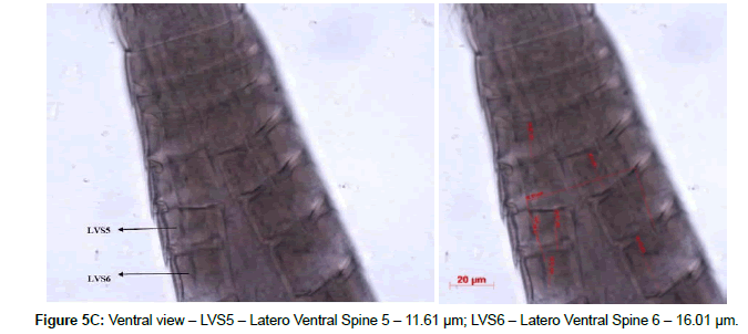
S5 Segment 5 (S5) had a length of 18.47 μm and its latero ventral spine show a length of 11.61 μm.The pachycyclus, sternal and tergal plate are similar to preceding one and middorsal spine had a length of 22.29 μm.
S6 Segment 6 (S6) showed a length of 28.16 μm with a 16.01 μm length of latero ventral spine.The pachycyclus, sternal and tergal plate are similar to preceding one and middorsal spine had a length of 20.39 μm.
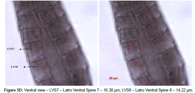
S7 Segment 7 (S7) exhibited a length of 27.54 μm with a 16.30 μm length of latero ventral spine,The pachycyclus, sternal and tergal plate similar to preceding one and the middorsal spine length was 20.64 μm.
S8 Segment 8 (S8) had a length of 24.04 μm with a latero ventral spine, which had a length of 14.22 μm. The pachycyclus, sternal and tergal plate are similar to preceding one and the middorsal spine has a length of 15.65 μm..
S9 Segment 9 (S9) exhibited a 26.72 μm length. The pachycyclus, sternal and tergal plate are similar to preceding of earlier one. The latero ventral spine present in this segment with a length of 12.64.
S10 Segment 10 (S10) had a length of 27.25 μm., The pachycyclus, sternal and tergal plate similar to preceding one, there were no middorsal and latero ventral spines.
S11 Segment 11 (S11) showed a length of 17.02 μm with a 112.29 μm length of lateral terminal spine.The percentage of LTS with TL is 43.19% One pair of lateral terminal accessory spine (LTAS) 11.02 μm long. Further, there were no latero ventral spine and middorsal spine. Terminal sternal plate slightly pointed.
Based on the external morphology the studied specimen was identified as an adult female.
Classification
Phylum: Kinorhyncha [4]
Class: Cyclorhagida [44]
Order: Echinorhagata [44]
Family: Echinoderidae (Bütschli, 1876)
Genus: Echinoderes (Claparède, 1863)
Species: Echinoderes remanei [64]
This specimen deposited in the Department of Ocean Studies and Marine Biology, Pondicherry University, Port Blair (DOSMB, PU, PB) depository. The Specimen Number is: DOSMB 06005
Habitat: The intertidal sediments of Wandoor, South Andaman. Mid Tide environment represented this species. This specimen observed only in the study period of First Phase of Monosoon. One specimen was collected.
Distribution: This species was identified in the North West Atlantic Ocean [64], Buzzards Bay, Massachusetts [10], San Juan Island [16], North of Cape Hatteras [19] and Gulf of Mexico [65], The present study reported from Wandoor (Lat.11°35’10.74”N – Long.092°37’1328”E), South Andaman Islands, India. Only one specimen was collected.
Discussion: The Echinoderes remanei species identified by Higgins [16] reported that the total length has 282–358 μM, however, the present studied species reported that the total length had 260 μM, which was lesser than the prescribed length suggested that it may be inferred that this species not matured enough.
Echinoderes truncates (Figures 6A-6F)
Diagnosis: This specimen’s head consists of the retractable mouth cone and oral stylets are present, but their exact number and arrangement could not be determined due to submerged in the mounting media. Head and Neck region are not considered as a segment. Total Trunk Length (TL) is 410.29 μm and Mean Sternal Width (MSW-6) is 99.09 μm. The percentage of MSW-6 with TL is 24.16%. The Standard Width (SW-10) is 78.06 μm and has the percentage with TL is 19.75%. Trichoscalid, spinoscalid plates and outer oral stylet are present, but it is not clearly visible. Midventral placid wider (25.02 μm) and other placid are smaller (13.59 μm). The trunk transverse section shows a triangular shape. Pectinate fringes moderately and visible to entire segment. Mid-terminal spine absent and first two trunk segment consist of a closed cuticular ring. Segment 6-9 exhibited latero ventral spines. Muscle scars present in the segments 3-10 ventral side.
Description
S1 Segment 1 (S1) showed a length 37.03 μm and closed cuticular ring visible. The pachycyclus well developed. There is no middorsal spine and sensory spots on middorsal position. Cuticular hair present.
S2 Segment 2 (S2) had a length of 40.05 μm. The pachycyclus well developed. The cuticular ring was closed on ventral side of the specimen and not shows any middorsal spine. The small round shaped cuticular hair present.
S3 Segment 3 (S3) exhibited a length of 42.04 μm, with two sternal plates, in ventral side and one tergal plate on the dorsal side. The pachycyclus similar to preceding one, there is no middorsal spine. Cuticular hair present.
S4 Segment 4 (S4) was having a length of 44.79 μm. The pachycyclus, sternal and tergal plate are similar to preceding one and the middorsal spine has a length of 22.87 μm. There is no latero ventral spine. One pair of scare present on dorsal side of the specimen. Cuticular hair present.
S5 Segment 5 (S5) has a length of 52.99 μm with the middorsal spine of 22.2 μm length present. The pachycyclus, sternal and tergal plate are similar to preceding one and the latero ventral spine present with a length of 11.05 μm. Cuticular hair present.
S6 Segment 6 (S6) showed a length of 52.88 μm with a latero ventral spine which has the 18.21 μm. The middorsal spine with a length of 10.97 μm present. The pachycyclus, sternal and tergal plate are similar to preceding one. Cuticular hair present.
S7 Segment 7 (S7) exhibited a length of 54.98 μm and with a latero ventral spine with a length of 18.23 μm. The middorsal spine has a length of 21.13 μm. The pachycyclus, sternal and tergal plate similar to preceding one and sensory spots absent. One pair glandular cell outlet, Type II present in dorsal side. Cuticular hair present.
S8 Segment 8 (S8) has a length of 50.07 μm with a length of 19.15 latero ventral spine present. The pachycyclus, sternal and tergal plate are similar to preceding one and 15.05 μm lengths of middorsal spine present. Sensory spots absent. One pair glandular cell outlet, Type II present in dorsal side. Cuticular hair present.
S9 Segment 9 (S9) exhibited a 51.14 μm length with 17.74 μm lengths of latero ventral spine. The pachycyclus, sternal and tergal plate are similar to preceding one and middorsal spine was absent. Sensory spots were absent. One pair glandular cell outlet, Type II present in dorsal side. Cuticular hair present.
S10 Segment 10 (S10) had a length of 47.9 μm, The pachycyclus, sternal and tergal plate similar to preceding one. The middorsal and lateral spines are absent. One pair of sensory spot present on ventral side of the specimen. Cuticular hair present.
S11 Segment 11 (S11) showed a length of 41.00 μm with a 176.49 μm length of lateral terminal spine (LTS) . The percentage of LTS with TL is 43.01%. Three pairs of penile spine with a length of (P1) 27.74 μm, (P2) 16.43 μm and (P3) 25.04 μm were observed. Further, there are no lateral spine and middorsal spine present. Terminal sternal slightly truncated. Cuticular hair present.
Based on the external morphology the studied specimen was identified as an adult male.
Classification
Phylum: Kinorhyncha [4]
Class: Cyclorhagida [44]
Order: Echinorhagata [44]
Family: Echinoderidae (Bütschli, 1876)
Genus: Echinoderes (Claparède, 1863)
Species: Echinoderes truncatus [19]
This specimen deposited in the Department of Ocean Studies and Marine Biology, Pondicherry University, Port Blair (DOSMB, PU, PB) depository. The Specimen Number is: DOSMB 06006
Habitat: The intertidal sediments of Dollygunj, South Andaman. Mid Tide environment represented this species. This specimen observed only in the study period of Second Phase of Monosoon. One specimen was collected.
Distribution: This species was identified in the Twin Cays, Atlantic Barrier Reef [19], Disco Island, West Greenland [52], Bocas del Tora, Panama [61] and Florida [62], The present study reported from Dollygunj (Lat.11°38’14.05”N – Long.092°42’10.80”E) South Andaman Islands, India. Only one specimen was collected.
Echinoderes bengalensis (Figure 7)
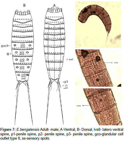
Diagnosis: This specimen’s head consists of the retractable mouth cone and oral stylets are present, but their exact number and arrangement could not be determined due to submergence in the mounting media. The head and neck regions were not considered as a segment. The total Trunk Length (TL) was 305 μm and Mean Sternal Width (MSW-6) was 59.37 μm long. The percentage of MSW-6 with reference to TL was 19.46. The Standard Width (SW-10) was 41.06 μm and its percentage with TL was 13.46. The trichoscalid, spinoscalid plates and outer oral stylet were present, but it is not clearly visible. Midventral placid wider (11.91 μm) and other placid were small (6.18 μm). Trunk transverse section showed a triangular shape. Pectinate fringes moderately and visible to entire segment. Mid-terminal spine and middorsal spine were absent. The first two trunk segments were closed and the cuticular ring was observed. Segment 5 and 8 exhibited latero ventral spines.
Description:
S1 Segment 1 (S1) showed a length of 16.87 μm and closed in nature. The cuticular ring was visible and the pachycyclus were well developed. There was no middorsal spine and latero ventral spines.
S2 Segment 2 (S2) showed a length of 21.16 μm and the well developed pachycyclus. The cuticular ring was closed on ventral side of the specimen and not showed any middorsal spine and latero ventral spines.
S3 Segment 3 (S3) exhibited a length of 22.59 μm with two sternal plate in ventral side and one tergal plate in dorsal side. The pachycyclus were similar to preceding segment. There were no middorsal spine and latero ventral spines.
S4 Segment 4 (S4) had a length of 22.71 μm. The pachycyclus, sternal and tergal plate were similar to preceding segment. There were no mid-dorsal, latero ventral spine and sensory spot.
S5 Segment 5 (S5) had a length of 27.89 μm with a latero ventral spine had a length 0f 10.05 μm. The pachycyclus, sternal and tergal plate were similar to preceding segment and there was no mid-dorsal, latero ventral spine and sensory spot.
S6 Segment 6 (S6) showed a length of 33.17 μm. The pachycyclus, sternal and tergal plate are similar to preceding segment and middorsal spine and latero ventral spine were absent. One pair of glandular cell outlet, Type II present in latero dorsal side .two pair of sensory spots presents on middorsal position.
S7 Segment 7 (S7) exhibited a length of 39.57 μm. The pachycyclus, sternal and tergal plate were similar to preceding segment. The middorsal spine was absent. One pair of glandular cell outlet, Type II present in latero dorsal and two pair of sensory spots present on middorsal position.
S8 Segment 8 (S8) had a length of 41.25 μm with a 10.06 μm long latero ventral spine . The pachycyclus, sternal and tergal plate were similar to preceding one and middorsal spine absent. One pair of glandular cell outlet type II present in latero dorsal and two pair of sensory spots present on middorsal position.
S9 Segment 9 (S9) exhibited a 50.74 μm. The pachycyclus, sternal and tergal plate were similar to preceding one and middorsal spine absent. One pair of glandular cell outlet, Type II and scare present on latero dorsal side. Two pairs of sensory spots present on middorsal position.
S10 Segment 10 (S10) had a length of 22.49 μm, The pachycyclus, sternal and tergal plate similar to preceding segment was observed. The mid dorsal, lateral spines and glandular cell outlet type II were absent. Three pair of sensory spots were present on middorsal position.
S11 Segment 11 (S11) showed a length of 14.35 μm with a 108.13 μm long lateral terminal spine (LTS) showed a percentage of 35.4 with reference to TL. Two pairs of penile spine with a length of (P1) 14.48 μm, (P2) 11.41 μm there is no P3. Further, there are no Lateral spine and middorsal spine. Terminal sternal slightly pointed.
Based on the external morphology the studied specimen was identified as an adult male.
Classification
Phylum: Kinorhyncha [4]
Class: Cyclorhagida [44]
Order: Echinorhagata [44]
Family: Echinoderidae (Bütschli, 1876)
Genus: Echinoderes (Claparède, 1863)
Species: Echinoderes bengalensis
This specimen deposited in the Department of Ocean Studies and Marine Biology, Pondicherry University, Port Blair (DOSMB, PU, PB) depository. The Specimen Number is: DOSMB 06007
Habitat: The intertidal sediments of Dollygunj and Carbyns Cove, South Andaman. Mid and Low Tidal environment represented this species. This specimen observed only in the study period of the Summer and First Phase of Monsoon. Three specimens were collected.
Distribution: This species was identified in Waltaier [66], Bangladesh [67], and Ganges Delta Area [19], The present study reported from Dollygunj (Lat.11°38’14.05”N – Long.092°42’10.80”E) and Carbyns Cove (Lat.11°38’20.25”N – Long.092°44’54.24”E) South Andaman Islands, India. Only three specimens were collected.
Discussion: The Echinoderes bengalensis samples were collected from the intertidal zone [1,19,21] was support the present study collection environment.
Discussion
The identified kinorhyncha species fall under the single environment called as intertidal environment. The species Echinoderes capitatus, Echinoderes peterseni, Echinoderes horni, Echinoderes remanei were observed in First Phase of Monsoon. Echinoderes truncatus represented Second Phase of Monsoon and Echinoderes bengalensis observed in First Phase of Monsoon to Summer. However, the species Echinoderes peterseni, eventhough it was reported in Arctic region, it has been identified in this tropical region is considered as mystery or some factors which are affecting was not able to identified in this part. Over and above, some temperatate speceis was reported from this part of waters support the possibilities of existence of this species in this tropical region. The species However, a constant monitoring and more elaborate work should be carried out in this aspect.
Conclusion
This research article narrates the status of all the identified Kinorhyncha species. A total of nine specimens encountered during the study period for these six species. Out of six species five (E. truncates, E. capitatus, E. remanei, E. horni, E. peterseni) species are considered as a new report to India and one species (E. bengalensis) reported after 38 years of its publication from Indian waters and new reports from Andaman waters. Since, there is not much work on Kinorhyncha studies in India, this work helps to initiate further research on this group.
Acknowledgements
The authors thank Dr. Hiroshi Yamasaki (Japan) for his tireless help and suggestions. They also acknowledge The Head, Department of Ocean Studies and Marine Biology and authorities of Pondicherry University for providing the facilities to execute this project.
References
- Neuhaus B (2013) Kinorhyncha (= Echinodera): Handbook of Zoology- Gastrotricha, Cycloneuralia and Gnathifera: Nematomorpha, Priapulida, Kinorhyncha, Loricifera. (1st edtn), Walter de Gruyter, Berlin, Germany.
- Horn TD (1978) The Distribution of Echinoderes coulli (Kinorhyncha) along an Interstitial Salinity Gradient. Trans Am Microsc Soc 97: 586-589.
- Modig H, Olafsson E (1998) Responses of Baltic benthic invertebrates to hypoxic events. J Exp Mar Bio Ecol 229: 133-148.
- Dujardin F (1851) Observations zoologies I. On a small marine animal, Echinodère, forming an intermediate type between crustaceans and worms. Ann Nat Sci Zoo Ser 15: 158-160.
- Reinhard Z (1885) Kinorhyncha (Echinoderes), their anatomical structure and their place in the system. Works of the Society of Naturalists at the Imperial University of Kharkow 19: 205-305.
- Reinhard W (1887) Kinorhyncha (Echinoderes), its anatomical structure and its position in the system. J Sci Zool 45: 401-467.
- Zelinka C (1928) Monograph der Echinodera. Wilhelm Engelmann, Leipzig, Germany.
- Higgins RP (1960) A new species of Echinoderes (Kinorhyncha) from Puget Sound. Trans Am Microsc Soc 79: 85-91.
- Higgins RP (1961) Three New Homalorhagid Kinorhynchs from the San Juan Archipelago, Washington. J Elisha Mitchell Sci Soc Chapel Hill N C 77: 81-88.
- Higgins RP (1964) Three new kinorhyncha from the North Carolina coast. Bull Mar Sci 14: 479-493.
- Higgins RP (1965) The homalorhagid Kinorhyncha of Northeastern U. S. Coastal Waters. Trans Am Microsc Soc 84: 65-72.
- Higgins RP (1966) Echinoderes arlis, a new kinorhynch from the Arctic ocean. Pacific Sci 20: 518-520.
- Higgins RP (1967) The Kinorhyncha of New-Calcdunia. In Expedition Francaise sur rÄÂÃ?Â?cÄ©fs coralliens de la Nouvelle Caledonia. Editions of the Singcr-Polignac Foundation 2: 75-90.
- Higgins RP (1968) Taxonomy and postembryonic development of the Cryptorhagae, a new suborder for the mesopsammic kinorhynch genus Cateria. Trans Am Microsc Soc 87: 21-39.
- Higgins RP (1969) Indian Ocean Kinorhyncha: 1, Condyloderes and Sphenoderes, new cyclorhagid genera. Smithson Contrib Zool 14: 1-13.
- Higgins RP (1977) Two new species of Echinoderes (Kinorhyncha) from South Carolina. Trans Am Microsc Soc 96: 340-354.
- Higgins RP (1978) Echinoderes gerardi n.sp. and E. riedli (Kinorhyncha) from the Gulf of Tunis. Trans Am Microsc Soc 97: 171-180.
- Higgins RP (1982) Three New Species of Kinorhyncha from Bermuda. Trans Am Microsc Soc 104: 305-316
- Higgins RP (1983) The Atlantic Barrier Reef Ecosystem at Carrie Bow Cay, Belize, II: Kinorhyncha. Smithsonian Institution Press, Washington DC, USA.
- Higgins RP (1985) The Genus Echinoderes (Kinorhyncha: Cyclorhagida) from the English Channel. J Mar Biol Assoc U.K. 65: 785-800.
- Higgins RP (1986) A New Species of Echinoderes (Kinorhyncha: Cyclorhagida) from a Coarse-sand California Beach. Trans Am Microsc Soc 105: 266-273.
- Higgins RP (1990) Zelinkaderidae, a new family of cyclorhagid Kinorhyncha. Smithson Contrib Zool 500: 1-26.
- Higgins RP (1991) Pycnophyes chukchiensis, a new homalorhagid kinorhynch from the Arctic Sea. Proc Biol Soc Wash 104: 184-188.
- Adrianov AV, Higgins RP (1996) Pycnophyes parasanmansis, a new kinorhynch (Kinorhyncha. Homalorhagida Pycnophyidae) from San Juan Island, Washington. Proc Biol Soc Wash 109: 236-247.
- Pardos F, Higgins RP, Benito J (1998) Two new Echinoderes (Kinorhyncha, Cyclorhagida) from Spain, including a reevaluation of kinorhynch taxonomiccharacters. Zool Anz 237: 195-208.
- Martorelli S, Higgins RP (2004) Kinorhyncha from the stomach of the shrimp Pleoticus muelleri (Bate, 1888) from Comodoro Rivadavia, Argentina. Zool Anz 243: 85-98.
- Neuhaus B, Pardos F, Sørensen MV, Higgins RP (2013) Redescription, morphology, and biogeography of Centroderes spinosus (Reinhard, 1881) (Kinorhyncha: Cyclorhagida). Cah Biol Mar 54: 109-131.
- Neuhaus B, Blasche T (2006) Fissuroderes, a new genus of Kmorhyncha (Cyclorhagida) from the deep sea and continental shelf of New Zealand and from the continental shelf of Costa Rica. Zool Anz 245: 19-52.
- Sørensen MV (2007) A new species of Antygomonas (Kinorhyncha: Cyclorhagida) from the Atlantic coast of Florida, USA. Cah Biol Mar 48: 155-168.
- Sørensen MV, Heiner I, Ziemer O, Neuhaus B (2007) Tubulideres seminoli gen. et sp. nov. and Zelinkaderes brightae sp. nov. (Kinorhyncha, Cyclorhagida) from Florida. Helgol Mar Res 61: 247-265.
- Sørensen MV, Heiner I, Hansen JG (2009) A comparative morphological study of the kinorhynch genera Antygomonasand Semnoderes (Kinorhyncha: Cyclorhagida). Helgol Mar Res 63: 129-147.
- Sørensen MV, Accogli G, Hansen JG (2010) Postembryonic development of Antygomonas incomitata (Kinorhyncha: Cyclorhagida). J Morphol 271: 863-882.
- Sørensen MV, Rho HS, Min WG, Kim D, Chang CY (2012) An exploration of Echinoderes (Kinorhyncha: Cyclorhagida) in Korean and neighboring waters, with the description of four new species and a redescription of E.tchefouensis Lou, 1934. Zootaxa 3368: 161-196.
- Sánchez N, Pardos F, Herranz M, Benito J (2011) Pycnophyes dolichurus sp. nov. and P. aulacodes sp. nov. (Kinorhyncha, Homalorhagida, Pycnophyidae), two new kinorhynchs from Spain with a reevaluation of homalorhagid taxonomic characters. Helgol Mar Res 65: 319-334.
- Sánchez N, Herranz M, Benito J, Pardos F (2012) Kinorhyncha from the Iberian Peninsula: new data from the first intensive sampling campaigns. Zootaxa 3402: 24-44.
- Sánchez N, Herranz M, Benito J, Pardos F (2014) Pycnophyes almansae sp. nov. and Pycnophyes lageria sp. nov., two new homalorhagid kinorhynchs (Kinorhyncha, Homalorhagida) from the Iberian Peninsula, with special focus on introvert features. Mar Biol Res10: 17-36.
- Sánchez N, García-Herrero A, García-Gómez G, Pardos F (2017) A new species of the recently established genus Setaphyes (Kinorhyncha, Allomalorhagida) from the Mediterranean with an identification key. Mar Biodivers 48: 249-258.
- Sørensen MV (2014) First account of echinoderid kinorhynchs from Brazil, with the description of three new species. Mar Biodivers 4: 251-274.
- Yamasaki H, Fujimoto S (2014) Two new species in the Echinoderes coulli group (Echinoderidae, Cyclorhagida, Kinorhyncha) from the Ryukyu Islands, Japan. ZooKeys 382: 27-52.
- Zotto MD (2015) Antygomonas caeciliae, a new kinorhynch from the Mediterranean Sea, with report of mitochondrial genetic data for the phylum. Mar Biol Res 11: 689-702.
- Landers SC, Sørensen MV (2016) Two new species of Echinoderes (Kinorhyncha, Cyclorhagida), E. romanoi sp. n. and E. joyceae sp. n., from the Gulf of Mexico. Zoo Keys 594: 51-71.
- Sørensen MV (2013) Phylum Kinorhyncha. Zootaxa. 3703: 063-066.
- Yamasaki H, Hiruta SF, Kajihara H (2013) Molecular phylogeny of kinorhynchs. Mol Phylogenet Evol 67: 303-310.
- Sørensen MV, Zotto DM, Rho HS, Herranz M, Sánchez N, et al. (2015) Phylogeny of Kinorhyncha Based on Morphology and Two Molecular Loci. PLoS One 10: e0133440.
- Rao GC, Ganapati PN (1966) Occurrence of an Aberrant Kinorhynch Cateria styx Gerlach, in Waltair Beach Sands. Curr Sci 35: 212-213.
- Rao GC, Ganapati PN (1962) Ecology of the Interstitial Fauna Inhabiting the Sandy Beaches of Waltair Coast. J Mar Biol Ass India 4: 44-57.
- Dovgal I, Chatterjee T, Ingole B, Nanajkar M (2008) First report of Limnoricus ponticus Dovgal & Lozowskiy (Ciliophora: Suctorea) as epibionts on Pycnophyes (Kinorhyncha) from the Indian Ocean with key to species of the genus Limnoricus. Cah Biol Mar 49: 381-385.
- Venkataraman K, Raghunanthan C, Sivaleela G, Choudury S, Mondal T, et al (2015) Lesser known marine animals of India. Zoological Survey of India, Kolkata, India.
- Greeff R (1869) Investigations on some strange groups of animals of the arthropod and worm type. Arch Nat Hist 35: 71-121.
- Jeeva C, Mohan PM (2016) A Report of Echinoderes setiger Greeff, 1869 (Kinorhyncha, Cyclorhagida) in Intertidal Zone of Port Blair. J Andaman Sci Assoc 21: 205-256.
- Zelinka C (1907) To the knowledge of the Echinoderen. Zool Anz 32: 130-136.
- Higgins RP, Thiel H (1988) Introduction to the study of meiofauna. Smithsonian Institution Press, Washington DC,
- Giere O (2009) The Microscopic Motile Fauna of Aquatic Sediments: Meiobenthology. (2nd edtn), Springer-Verlag, Berlin, Germany.
- Pardos MV, Herranz M, Landers SC (2016) A new species of Echinoderes (Kinorhyncha: Cyclorhagida) from the Gulf of Mexico, with a redescription of Echinoderes bookhouti Higgins, 1964. Zool Anz J Comp Zool 265: 48-68.
- Hyus R, Coomans A (1989) Echinoderes higginsi sp.n. (Kinorhyncha, Cyclohagida) from the southern North Sea with a key to the genus Echinoderes Calparede. Zool Scr 18: 211-221.
- Higgins RP, Kristensen RM (1988) Kinorhyncha from Disko Island, West Greenland. Smithson Contrib Zool 458: 1-56.
- Grzelak K, Sorensen MV (2017) New species of Echinoderes (Kinorhyncha: Cyclorhagida) from Spitsbergen, with additional information about known Arctic species. Mar Biol Res 14: 113-147.
- Sachithanandam V, Mohan PM, Karthik R, Saielangovan S, Padmavati G (2013) Climate changes influence the phytoplankton bloom (prymnesiophyceae: Phaeocystis spp.) in North Andaman coastal region. Indian J Geomarine Sci 42: 58-66.
- Mohan PM, Muruganantham M, Ubare VV (2017) A Report of Uncommon Sabellariid Polychaete Worm to Tropical Island Environment of Andaman Archipelago, Andaman Sea. J Mar Biol Oceanogr 6:3.
- Jhimli M (2018) Studies on reff associated ascidians of Andamn and Nicobar Islands, India. PhD Thesis Pondicherry University, Puducherry, India.
- Zotto MD, Domenico MD, Garraffoni A, Sørensen MV (2013) Franciscideres gen. nov. – a new, highly aberrant kinorhynch genus from Brazil, with an analysis of its phylogenetic position. Syst Biodivers 11: 303-321.
- Pardos F, Sánchez N, Herranz M (2016) Two sides of a coin: The Phylum Kinorhyncha in Panama. I) Caribbean Panama. Zool Anz 265: 3-25.
- Herranz H, Boyle MJ, Pardos F, Neves RC (2014) Comparative myoanatomy of Echinoderes (Kinorhyncha): a comprehensive investigation by CLSM and 3D reconstruction. Front Zool 11: 01-26.
- Blake CH (1930) Three new species of worms belonging to the order Echinodera. Biological Survey Mount Desert Is. Region Part 4: 3-10.
- Landers SC, Sørensen MV, Beatona KR, Jonesa CM, Millera JM, et al. (2017) Kinorhynch assemblages in the Gulf of Mexico continental shelf collected during a two-year survey. J Exp Mar Bio Ecol 502: 81-89.
- Higgins RP, Rao GC (1979) Kinorhynchs from the Andaman Islands. Zool J Linn Soc 67: 75-85.
- Timm RW (1958) Two new species of Echinoderiderella (Phylum Kinorhyncha) from the Bay of Bengal. J Bombay Nat Hist Soc 55: 107-109.
 Spanish
  Chinese
  Russian
  German
  French
  Japanese
  Portuguese
  Hindi
Hindi 


