Research Article, J Clin Exp Onco Vol: 12 Issue: 4
In vitro Study-Anti-Cancer Activity of Conium Maculatum in MCF-7 Breast Cancer Cell Line
Dr. Jeba Beula*
1Department of Materia Medica, White Memorial Homeopathic Medical College, Attoor, Tamil Nadu
*Corresponding Author: Dr. Jeba Beula,
Department of Materia Medica, White
Memorial Homeopathic Medical College, Attoor, Tamil Nadu
E-mail: drpnisha15@gmail.com
Received: 12 July, 2023, Manuscript No. JCEOG-23-105826
Editor assigned: 17 July, 2023, PreQC No. JCEOG-23-105826 (PQ);
Reviewed: 01 August, 2023, QC No. JCEOG-23-105826;
Revised: 09 August, 2023, Manuscript No. JCEOG-23-105826 (R);
Published: 17 August, 2023 DOI: 10.4172/2324-9110.1000360
Citation: Beula J (2023) In vitro Study-Anti-Cancer Activity of Conium Maculatum in MCF-7 Breast Cancer Cell Line. J Clin Exp Oncol 12:4.
Abstract
Background: Breast cancer a major cause of mortality worldwide. Although the disease occurs all over the world, its mortality, morbidity and survival rates vary based on etiological
factors. In spite there are various several modern treatments for breast cancer, there is a high risk for recurrence in majority of treatments. Hence, this study aimed to investigate the
efficacy of homoeopathic medicine Conium maculatum 6CH and 200CH in Michigan Cancer Foundation-7 (MCF-7) breast cancer cell line.
Methods: Assessed the cell viability by Mean Transit Time (MTT) assay. Cytotoxic effects of Conium maculatum by Sulforhodamine B (SRB) assay and Cell proliferation using colony forming assay.
Results: Ultra diluted preparation of the homoeopathic medicine Conium maculatum has significant effects in specific breast cancer cell line, producing cytotoxicity and a decrease in
cell proliferation.
Conclusion: It is scientifically evident that the homoeopathic medicine Conium maculatum has a significant effect in breast cancer cell line
Keywords: Breast cancer; Conium maculatum; Cytotoxicity; Homoeopathy; MCF7 cell lines
Abstract
Background: Breast cancer a major cause of mortality worldwide. Although the disease occurs all over the world, its mortality, morbidity and survival rates vary based on etiological factors. In spite there are various several modern treatments for breast cancer, there is a high risk for recurrence in majority of treatments. Hence, this study aimed to investigate the efficacy of homoeopathic medicine Conium maculatum 6CH and 200CH in Michigan Cancer Foundation-7 (MCF-7) breast cancer cell line.
Methods: Assessed the cell viability by Mean Transit Time (MTT) assay. Cytotoxic effects of Conium maculatum by Sulforhodamine B (SRB) assay and Cell proliferation using colony forming assay.
Results: Ultra diluted preparation of the homoeopathic medicine Conium maculatum has significant effects in specific breast cancer cell line, producing cytotoxicity and a decrease in cell proliferation.
Conclusion: It is scientifically evident that the homoeopathic medicine Conium maculatum has a significant effect in breast cancer cell line.
Keywords
Breast cancer; Conium maculatum; Cytotoxicity; Homoeopathy; MCF7 cell lines
Introduction
With reference to World Health Organization (WHO), increasing breast cancer outcomes and survival through early detection remains a cornerstone of breast cancer conventions. Breast cancer is treated using a variety of modern treatments including targeted therapy, hormone therapy, radiation therapy, surgery, and chemotherapy. Individuals with metastasis characteristically aimed at alleviation of symptoms and prolongation of survival. The major hostile side effects of breast cancer treatment are one of the most motivating factors to find some alternative methods [1-4].
Epidemiology
Breast cancer is the second most cause of death (1.38 million, 10.9% of all cancer) worldwide after lung cancer [5]. According to Global Cancer Observatory (GLOBOCAN), this is the most common cancer among women, accounting for 25.1% of all the cancers. Breast cancer incidence is higher in developed countries, whereas relative mortality is greatest in less developed countries [6]. Worldwide, breast cancer comprises 10.4% of all cancer incidences among women and about 100 times more common than women [7]. A study conducted by GLOBOCAN an international agency for research and WHO in the year 2020 on cancer, states that the incidence and mortality caused by the breast cancer accounts for 2.3 million. Among new cases (11.6%) the mortality rate of female breast cancer estimates to be 685,000 (6.9%) of all cancers. The study carried out by National Breast Cancer Foundation, United States 2020, reveals that 1 in 8 women may develop breast cancer in their life time among them the incidence of invasive breast cancer is found to be 276,480 in new cases as well as non-invasive (in situ) accounts for about 48,530 in new cases.
Conium maculatum, an extremely poisonous flowering weed, known as Hemlock which belongs to the family Apiacea, comprises several pyridine alkaloids like coniine, N-methylconiine, conhydrine, pseudoconhydrine and gamma-coniceine, precursors of some other hemlock alkaloids [8]. This plant is highly toxic in nature and its extract has been used as a traditional remedy for different diseases in olden days [9]. In homeopathy, it is used as a remedy for breast, cervix and uterine cancer [10]. Since Conium is a rich in alkaloids which have been reported that have anti-cancer properties [11-13]. Conium acting on the glandular system, we naturally expect it to be a great anti-scrofulous and anti-cancerous remedy. Cell lines play an important role in studying physiological, pathophysiological, and differentiation processes of specific cells. It allows the examination of stepwise alteration in its structure, biology, and genetic makeup of the cell under controlled environments [14,15]. Anti-cancer potential of Conium maculatum extract against cancer cells in vitro was explored by Mondal et al., It has the potential to interact with the DNA and hinders the process of cell proliferation and cell cycle. And also it reduces the cell viability and colony formation ability of HeLa cells; reduce cell proliferation and cell cycle arrest [16,17].
Materials and Methods
Study setting
This study was designed to evaluate the effectiveness of homoeopathic medicine Conium maculatum in breast cancer cells.
Intervention: Conium maculatum 6CH and 200CH dilutions prepared from Good Manufacturing Practices (GMP) certified Wilmer Schwabe Company.
Cell culture: The breast cancer cell line namely MCF-7 was purchased from National Centre for Cell Science (NCCS), Pune, India. The cells were grown in Dulbecco's Modified Eagle Medium (DMEM) supplemented with 10% Fasting Blood Sugar (FBS), nonessential amino acids and penicillin and streptomycin at 1X final concentration from a 100X stock and were maintained in a Carbon Dioxide (CO2) incubator at with 5% CO2 and 95% humidity atmosphere. Once the cells attained confluent growth, the cells were trypsin zed using Trypsin-Ethylene Diamine Tetra Acetic acid (EDTA) and the cells were seeded into sterile 6-well and 96-well plates for carrying out various assays. The cytotoxicity assays were carried out in 96-well plates and staining was performed in 6-well plates. In each well of the 6-well plates, a clean, dry, sterile coverslip was placed before the cells were seeded, followed by the incubation in a CO2 incubator (Innova CO-170, United States) with 5% CO2 and 95% humidity atmosphere.
Treatment groups:
1. Cells alone (Control)
2. Cells+Conium maculatum (6C)
3. Cells+Conium maculatum (200C)
Cell Growth Inhibition Studies
Cell viability assay–MTT assay
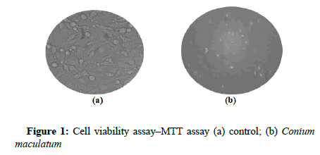
The viability of cells after concentration-and time-dependent treatments was determined using the standard MTT [3-(4,5- Dimethylthiazol-2-yl)-2,5-Diphenyltetrazolium Bromide] assay. Briefly, 100 μl of cells were seeded in 96 well plates at a density of 1×10 6 cells/ml and it was exposed to different concentrations of Conium maculatum (6C and 200C) ranging from 10 μl, 25 μl, 50 μl and 100 μl for 24 hours. After 24 hours’ treatment the cells were treated with 50 μl of MTT (Sigma, USA) and kept undisturbed for 3 hours at 37 ̊C. After the incubation period 200 μl of PBS was added to all the samples and the excess MTT was removed by aspiration. To that 200 μl of isopropanol was added and for solubilisation it was left overnight in the dark and the absorbance was read in micro titre plate at 560 nm (Biorad, iMark Microplate Reader) and the percentage viability of the treatment groups were calculated. This was done in triplicate with cell viability and the 50% of growth Inhibition Concentration (IC50) was calculated from a plotted dose-response curve (Figure 1). After fixing the dose with IC50 value, MTT assay was then repeated.
Sulphorodamine B ASAY – SRB
A portion of 350 μl of ice cold 40% Tricarboxylic Acid (TCA) was layered on top of the treated cells and incubated at 4 ̊C for one hour. The cells were then washed 5 times with 200 μl of cold PBS. The buffer was removed and 350 μl of SRB stain was added to each well and left in contact with the cells for 30 minutes at room temperature. The unbound dye was removed by washing four times with 350 μl portions of 1% acetic acid. Then 10 mM Tris (350 μl) was added to each tube, to stabilize the protein-bound dye and plate was shaking gently for 20 minutes. The Tris layer in each tube was transferred to a 96-well plate and the absorbance was read in a microliter plate reader (Biorad, iMark Microplate Reader) 492 nm. The cell survival on MCF-7 was recorded and the percentage viability was calculated.
Colony Forming Assay
Colony formation assay was performed in a 6-well plate. 1 x 10³ cells of MCF-7 were seeded onto 6-well sterile plates. Next day, the cells were treated with treatment groups. After cell adhesion, media was aspirated and fresh media was added and incubated for 7 days at 37 ̊C incubator supplied with 5% CO2. The colonies developed were fixed with glutaraldehyde, stained using crystal violet.
Results
Effect of homeopathic medicine Conium maculatum on the viability of breast cancer cell line
Several approaches were used to investigate the cell viability/ cytotoxicity of MCF-7 breast cancer cell lines. The MTT cell proliferation assay measures the cell proliferation rate and conversely, the reduction in cell viability when metabolic events lead to apoptosis or necrosis. The yellow compound MTT is reduced by mitochondrial dehydrogenase to the water insoluble blue formazan compound, depending on the viability of the cells. MCF-7 cells were exposed to different concentrations of Conium maculatum (6CH and 200CH) ranging from 10 μl, 25 μl, 50 μl and 100 μl for 24 hours and the cell viability was assessed using MTT assay. The IC50 value was found to be 25 μl and 50 μl 25 μl and 50 μl for Conium maculatum (6C and 200C) treated group with an optimal exposure period of 24 hours. (Table 1). The MTT assay showed that Conium maculatum significantly inhibited the viability of MCF-7 cells in a dose dependent manner. (Table 2).
| Volume of Sample | 6C | % Inhibition |
|---|---|---|
| 10 µl | 0.394 | 28.24 ± 0.274 |
| 25 µl | 0.257 | 53.18 ± 1.27 |
| 50 µl | 0.179 | 67.33 ± 0.64 |
| 100 µl | 0.113 | 79.45 ± 0.55 |
Table 1: Conium maculatum 6CH
| Volume of sample | 200C | % Inhibition |
|---|---|---|
| 10 µl | 0.434 | 21.08 ± 0.72 |
| 25 µl | 0.34 | 38.06 ± 0.1 |
| 50 µl | 0.267 | 51.45 ± 0.18 |
| 100 µl | 0.203 | 62.36 ± 0.62 |
Table 2: Conium maculatum 200CH
Sulforhodamine B (SRB) assay
SRB assay is the indicative of the metabolic capacity of the cells. Percent inhibition of metabolic activity has been measured and the control value has been fixed to be 100 percent of other groups which will be calculated relative to that. Anticancer efficacy of Conium maculatum was screened using MCF-7, breast cancer cell line at 6C and 200C concentrations. Extract inhibited the growth of the cells in dose dependent manner. The effect was found to be at 25 μl and 50 μl for Conium maculatum (6C and 200C) treated group with an optimal exposure period of 24 hours. (Table 3).
| Volume of sample | % Inhibition | |
|---|---|---|
| 6C | 200C | |
| 10 µl | 27.27 ± 0.29 | 23.18 ± 0.77 |
| 25 µl | 55.23 ± 1.33 | 36.22 ± 0.13 |
| 50 µl | 65.35 ± 0.74 | 52.48 ± 0.22 |
| 100 µl | 80.54 ± 0.59 | 61.88 ± 0.65 |
Table 3: Conium maculatum
Colony forming assay
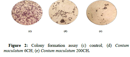
Clonogenic assay an in vitro cell survival assay based on the ability of a single cell to grow into a colony. We analyzed if the sample Conium maculatum could impair colony formation. The results showed that on exposure to the samples there was a decrease in the cell population and this indicates treatment group has an antiproliferative effect. (Figure 2).
Discussion
Breast cancer is a most common disease; instead of all modern treatment each person searches for anti-cancer agents from natural sources to reduce the side effects. Nowadays, Active constituents have been isolated and are used to treat breast cancer. So many studies were conducted in cancer cell lines using homoeopathic medicine like Jesmin model et al., about anti-cancer potential of Conium maculatum in HeLa cells of cervix [18]. Kaushik Bishayee et al., about effects of conium in HeLa cells [8]. Moshe frankel et al., about cytotoxicity of Conium and Phytolacca in MCF-7 and M231-MB-231 in breast cancer cell lines.
The present study examined the effects of Conium maculatum in breast cancer cells by using MTT Assay, SBB Assay and cell proliferation assay. Our findings suggest that ultra-diluted homoeopathic medicine Conium maculatum 6CH and 200CH exert preferential cytotoxic effects in MCF-7 breast cancer cell lines. This finding supports the findings of Kaushik Bishayee et al., IC50 was found to be 25 μl and 50 μl for Conium maculatum 6CH and 200 CH. SRB assay was done to assess the metabolic capacity of cells and this percent inhibition of metabolic activity is found to be 25 μl and 50 μl for Conium maculatum 6C and 200C. Also colony forming assay was done to assess the anti-proliferative effect. It shows a good decrease in cell population and has an anti-proliferative effect. The findings of the study correlates with the studies done by Jesmin model et al., and Moshe frankel et al., [18].
Conclusion
From results of present study, we can conclude that Conium maculatum hinders the process of cell proliferation and cell cycle and reduce the cell viability. The results of Conium maculatum on MCF-7 cell line showed that the sample had promising effects on inhibiting the cancer cells. The inhibitory effects observed on cell proliferation and cell cycle progression in the MCF-7 breast cancer cell line suggest that this plant extract may contain bioactive compounds with therapeutic potential. The exact mechanisms underlying its anticancer effects require further exploration, the present study provides a strong basis for conducting in-depth studies to identify and characterize the active compounds responsible for these inhibitory effects.
References
- Sainsbury JR, Anderson TJ, Morgan DA (2000) Breast cancer. Bmj 321(7263):745-750.
[Crossref]
- Akram M, Iqbal M, Daniyal M, Khan AU (2017) Awareness and current knowledge of breast cancer. Biol Res 50(1):1-23.
- Loscalzo (2022) Harrison’s principles of internal medicine, 17th edition, Volume II.
- Ghoncheh M, Mahdavifar N, Darvishi E, Salehiniya H (2016) Epidemiology, incidence and mortality of breast cancer in Asia. Asian Pac J Cancer Prev 17(3):47-52.
- Sharma GN, Dave R, Sanadya J, Sharma P, Sharma K, et al. (2010) Various types and management of breast cancer: an overview. J Adv Pharm Technol Res 1(2):109.
- Lopez TA, Cid MS, Bianchini ML (1999) Biochemistry of hemlock (Conium maculatum L.) alkaloids and their acute and chronic toxicity in livestock. A review. Toxicon 37(6):841-65.
- Boericke W (2004) Homeopathic materia Medica USA: Kessinger Publishing; 1:95–103.
- Bishayee K, Mukherjee A, Paul A, Khuda-Bukhsh AR (2012) Homeopathic mother tincture of Conium initiates reactive oxygen species mediated DNA damage and makes HeLa cells prone to apoptosis. Int J Genuine Tradit Med 2:37-41.
- Vetter J (2004) Poison hemlock (Conium maculatum L) Food Chem Toxico 42(9):1373-1382.
- Hussain AR, Ahmed SO, Ahmed M, Khan OS, Al Abdul Mohsen S, et al. (2012) Cross-talk between NFkB and the PI3-kinase/AKT pathway can be targeted in primary effusion lymphoma (PEL) cell lines for efficient apoptosis. Plosone 7(6):e39945.
- Singh BN, Shankar S, Srivastava RK (2011) Green tea catechin, epigallocatechin-3-gallate (EGCG): mechanisms, perspectives and clinical applications. Biochem Pharmacol 82(12):1807-21.
- Choudhuri NM (2014) A study on Materia Medica; B. jain publishers pvt ltd; 11th impression. 335-337.
- Russell RC, Williams NS, Bulstrode CJ, Bailey H, Love RJ, et al. (2000) Bailey and Love's short practice of surgery.
- Gey G (1952) Tissue culture studies of the proliferative capacity of cervical carcinoma and normal epithelium. Cancer Res 12:264-265.
- Igarashi M, Miyazawa T (2001) The growth inhibitory effect of conjugated linoleic acid on a human hepatoma cell line, HepG2, is induced by a change in fatty acid metabolism, but not the facilitation of lipid peroxidation in the cells. Biochim Biophys Acta 1530(2-3):162-71.
- Skehan P, Storeng R, Scudiero D, Monks A, McMahon J, et al. (1990) New colorimetric cytotoxicity assay for anticancer-drug screening. J Natl Cancer Inst 82(13):1107-12.
- Patrawala L, Calhoun-Davis T, Schneider-Broussard R, Tang DG (2007) Hierarchical organization of prostate cancer cells in xenograft tumors: the CD44+ α2β1+ cell population is enriched in tumor-initiating cells. Cancer Res.67(14):6796-6805.
- Mondal J, Panigrahi AK, Khuda-Bukhsh AR (2014) Anticancer potential of Conium maculatum extract against cancer cells in vitro: Drug-DNA interaction and its ability to induce apoptosis through ROS generation. Pharmacogn. Mag 10(3):524-533.
 Spanish
  Chinese
  Russian
  German
  French
  Japanese
  Portuguese
  Hindi
Hindi 


