Case Report, Int J Ophthalmic Pathol Vol: 10 Issue: 3
Basal Cell (Monomorphic) Adenoma of the Eyelid
Dokpe Emechebe2*, Majid Zeidi2, Ning Neil Chen2, Roman Shinder1,3
1Department of Ophthalmology, SUNY Downstate Medical Center, Brooklyn, New York United States
2Department of Pathology, SUNY Downstate Medical Center, Brooklyn, New York United States
3Department of Otolaryngology, SUNY Downstate Medical Center, Brooklyn, New York United States
- *Corresponding Author:
- Dokpe Emechebe
Department of Pathology
SUNY Downstate Medical Center, Brooklyn
New York, United States
E-mail: Dokpe.emechebe@downstate.edu
Received: March 01, 2021 Accepted: March 18, 2021 Published: March 25, 2021
Citation: Emechebe D, Zeidi M, Chen NN, Shinder R (2021) Basal Cell (Monomorphic) Adenoma of the Eyelid. Int J Ophthalmic Pathol 10:3.
Abstract
Basal Cell Adenoma (BCA) is an uncommon benign salivary gland epithelial neoplasm composed of basaloid cells lacking chondromyxoid stroma. BCA occurring in the eyelid is an extremely rare finding. We describe a case of BCA arising in the upper eyelid of a 30-year-old woman. A well-demarcated subcutaneous nodule arising in the eyelid was composed of islands of uniform basaloid cells arranged in tubular and trabecular patterns and ductal and myoepithelial cells surrounded by hyaline basement membrane material. Immunohistochemically, luminal cells stained positive for CK7 and CD117, myoepithelial cells stained with p63 and both luminal and myoepithelial cells stained with SOX10 confirming the diagnosis of BCA. Further pathological characteristics of this disease are discussed.
Keywords: Basal Cell Adenoma, Chondromyxoid stroma, pleomorphic adenoma, Eyelid
Keywords
Basal cell adenoma; Chondromyxoid stroma; Pleomorphic adenoma; Eyelid
Introduction
BCA is a benign tumor resembling pleomorphic adenoma, but with basaloid cells and peripheral palisading lacking chondromyxoid stroma. It accounts for 1 - 2% of epithelial tumors of salivary glands; 2% of benign salivary gland tumors. The most common involved site is the parotid gland; however other possible sites include the submandibular gland, minor salivary glands of upper lip, buccal mucosa, palate and nasal septum. Very few cases have been reported of BCA arising in the eyelid. The majority of the eyelid tumors, benign and malignant, are of cutaneous origin. Amongst the tumors arising from adnexal structures of the eyelid, pleomorphic adenoma is the most common.
Case Report
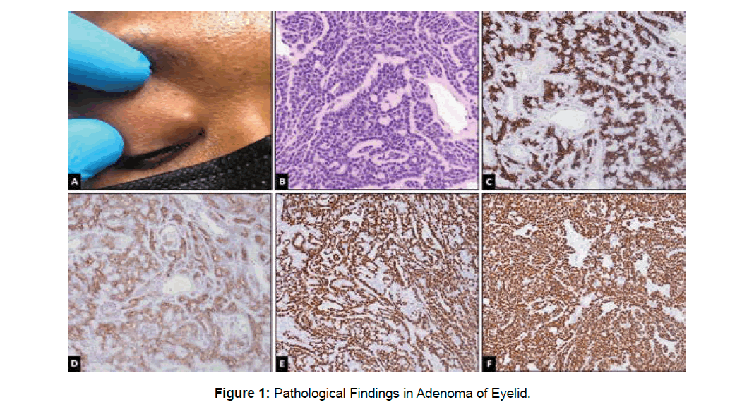
A 30-year-old African American woman was referred to our hospital for evaluation of a lesion of the right medial upper eyelid. The lesion has been present for several years and grew slowly over time. Physical examination revealed a well-defined, painless, and firm mass in the palpebrae interna of the middle of upper eyelid, (Figure 1A). The mucosa of the eyelid was normal, and no ulceration or induration was found. Other ophthalmic examination results were normal. The mass was easily dissected without any demonstrable involvement of the orbicularis oculi muscle or the wall of the canaliculus. The mass was excised and submitted for pathologic examination.
Pathological Findings
Microscopically, the lesion revealed islands of uniform basaloid cells arranged in tubular and trabecular patterns and ductal and myoepithelial cells surrounded by hyaline basement membrane material (Figure 1B). Luminal cells stained positive for CK7 (Figure 1C) and CD117 (Figure 1D), myoepithelial cells stained with p63 (Figure 1E), and both luminal and myoepithelial cells stained with SOX10 (Figure 1F) confirming a diagnosis of BCA.
Discussion
BCA is a rare benign epithelial tumor of the salivary gland, accounting for 1-2% of all salivary gland epithelial tumors [1]. On the basis of morphologic features BCA was divided into four subtypes: solid, tubular, trabecular, and membranous variants [2]. The most common type is the solid variant. According to previous studies of BCA, it accounts for 1-3% of all salivary gland tumors. The membranous variant of BCA has been reported in the eyelid [3] but the trabecular variant of BCA has not occurred in the eyelid reviewing the English literature. Histologically, BCA are classified as monomorphic tumors composed chiefly of basaloid cells organized with a prominent basal cell layer and distinct basement membrane-like material. The basaloid cells are sometimes indistinguishable from adenoid cystic carcinoma. Features such as: invasive growth, non-capsulated, myoepithelial cells constituted pseudo-glandular architectures, can help differentiate adenocystic carcinoma from BCA. Due to the rarity of such cases, some cutaneous origin tumors also should be ruled out. Basal cell carcinoma, for example, might have basal-like arrangements, and follicular originated tumors also have basal-like features. If we note that basal cell carcinoma is attached to the epidermis and usually has retraction artifact, follicular tumors have well-formed keratocysts or papillary mesenchymal bodies, and is associated with dermis, we can easily rule out such cutaneous origin tumors [4].
Conclusion
In general, our study encountered a rare case of trabecular variant BCA of the right upper eyelid. Though recently diagnosed, the 3 months of follow-up showed no recurrence detected in the case. Reviewing the literature, a case of malignant transformation of a BCA has been reported. Therefore, careful follow-up observation is important.
References
- Nagao K, Matsuzaki O, Saiga H, Sugano I, Shigematsu H et al. (1982) Histopathologic studies of basal cell adenoma of the parotid gland. Cancer 50:736–745.
- Araújo VC (2005) Basal cell adenoma In: Barnes L, Eveson JW, Reichart P, Sidransky D, editors. World Health Organization classification of tumors. Pathology and Genetics of the Head and Neck Tumours Lyon: IARC Press 261–262.
- Huang Y, Yang M, Ding J (2014). Membranous basal cell adenoma arising in the eyelid. Int J Clin Exp Pathol 7: 4508–4511.
- Nagao T, Sugano I, Ishida Y, Matsuzaki O, Konno A (1997) Carcinoma in basal cell adenoma of the parotid gland. Pathol Res Pract 193: 171–178
 Spanish
  Chinese
  Russian
  German
  French
  Japanese
  Portuguese
  Hindi
Hindi 
