Case Report, J Vet Sci Med Diagn Vol: 12 Issue: 2
An Interesting Case of a Linear Foreign Body in a Persian Cat
Maitha Al Muheiri, Khaja Mohteshamuddin*, Zaib Mahel and Azhar Ayub
Department of Veterinary Medicine, Arab Emirates University, Al Ain, United Arab Emirates
*Corresponding Author: Khaja Mohteshamuddin
Department of Veterinary
Medicine, Arab Emirates University, Al Ain, United Arab Emirates
E-mail: drkhaja707@uaeu.ac.ae
Received date: 31 October, 2022, Manuscript No. JVSMD-22-79016; Editor assigned date: 03 November, 2022, PreQC No. JVSMD-22-79016 (PQ); Reviewed date: 17 November, 2022, QC No. JVSMD-22-79016; Revised date: 09 February, 2023, Manuscript No. JVSMD-22-79016 (R); Published date: 16 February, 2023, DOI: 10.4172/2325-9590.100041
Citation: Muheiri MA, Mohteshamuddin K, Mahel Z, Ayub A (2023) An Interesting Case of a Linear Foreign Body in a Persian Cat. J Vet Sci Med Diagn 12:2
Abstract
Foreign bodies in the airways are a common problem in small animal practice, especially in domestic cats. A case report of recurrent ingested thread as a linear foreign body with the thread held back by the tongue and stuck causing further complications is presented in this case report. Interestingly, the radiographic results did not indicate the paisley shaped gas pattern which is a characteristic pattern of a linear foreign body. The final diagnosis was made by exploratory laparotomy which confirmed the presence of several threads in the small intestine. Therefore, this complex case was diagnosed based on history, clinical signs and results of radiography and confirmed by exploratory laparotomy. This is the first ever published case report of a linear foreign body in UAE according to the authors’ knowledge.
Keywords: Linear foreign body; Obstruction; Cat; Radiograph; Exploratory laparotomy
Introduction
Gastrointestinal foreign bodies are common in cats due to their high tendency to explore and eat any materials. Linear foreign body such as thread or string most often can be passed through with the stools, though it is possible for a small object to rattle around inside the stomach without passing for weeks. If the object does not pass and causes obstruction or partial obstruction, surgery will be required to remove it. Early diagnosis allows early removal of the foreign body before the bowel gets damaged [1]. Timely intervention will allow preventing complications such as peritonitis due to gastric or intestinal perforation [2]. However, some of these episodes might end up in fatal consequences.
The most common clinical signs of such condition described were persistent vomiting, lethargy, anorexia, depression, diarrhea, dehydration, painful abdomen, uncomfortable and an unusual hiding behaviour. Linear foreign body obstruction can result in chronic, intermittent, gastrointestinal disease in cats [3]. Acute or chronic obstruction may be combined with varying phases of dyspnoea, persistent coughing, haemoptysis, halitosis and similar symptoms associated with possible migration like pneumonia, pneumothorax, pulmonary abscess or pyothorax [4,5]. Tracheal and bronchial foreign bodies in the cat have been infrequently reported in the veterinary literature [6,7].
This case report describes the diagnosis and surgical intervention which led to an uneventful recovery of a Persian cat presented with gastrointestinal obstruction due to linear foreign body. This is the first ever published case report of linear foreign body in cats in the UAE according to the authors’ knowledge.
Case Presentation
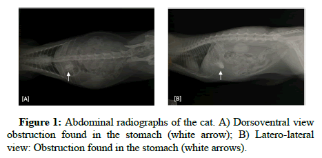
A 3 years old female persian cat named guccie was presented to paws and claws veterinary clinic for routine vaccination and deworming. The owner describes the cat as always being playful, active, living with many cats and very social. She is known for her obsession of eating threads but usually she gets rid of it either by vomiting or by defecating. Unfortunately, this time she was presented at the clinic with a history of swallowed threads, vomiting and anorexia. During the physical examination the patient appeared lethargic, her body weight was recorded as 3.5 kg with a normal body condition score and she was dehydrated with a pale mucus membrane. The body temperature was 39.5°C, evincing pain upon abdominal palpation, injured and swollen mouth due to the thread being held back by the tongue and it was unable to progress in the digestive tract. The patient was sedated immediately by inhalation anesthetics, the mouth was open and the thread was appearing to be wrapped around the tongue. The thread was identified and released. Unfortunately, after that we tried to remove it gently, but it was stuck somewhere inside, the patient was sent to the radiology room. Abdominal radiographs were taken to rule out other conditions (differential diagnosis). Upon radiographic examination, it indicated foreign bodies in the stomach (Figure 1). After five minutes the cat was a wake, vomited and get rid of the thread. After that the cat was sent to radiology room for further examination. Regarding the technique of positive contrast gastrography, the patient preparation is very important because the food can mimic a gastric lesion and therefore the patient should be fasted for 12-24 hours.
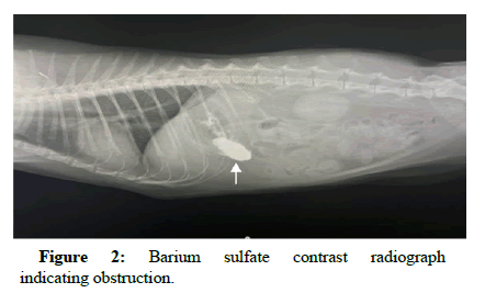
Usually, before proceeding with such a procedure, the animal should be fasting. However, in this case the patient was already anorexic in the last 2 days. The cat was sedated with isoflurane inhalation anesthetic and then barium sulfate was administered orally as a contrast agent. After that it has been recommended to gently turn the patient to achieve uniform coating of the gastric mucosa. Immediately after barium sulfate was administered, left lateral, right lateral, ventrodorsally and dorsoventral views of the stomach should be made, which are then usually followed by left recumbent lateral and ventrodorsally, views of the abdomen after 15 min, 30 min, 45 min and 1 h, 2 h, 3 h, 4 h or until complete emptying of the stomach content is present. This standard procedure was followed in this case as described above (Tables 1 and 2). The results indicated that the linear foreign body formed as an obstruction in the stomach because the barium sulfate did not pass by the stomach so the obstruction was in the first part of intestine (Figure 2). The cat was hospitalized for follow up, the day after the cat started to vomit with more intensity.
| Parameter | Result | Reference range | Unit |
|---|---|---|---|
| ALB | 2.9 | 2.2-4.4 | g/dL |
| ALP | 10 | 10-90 | U/L |
| ALT | 71 | 20-100 | U/L |
| TBIL | 419 | 300-1100 | U/L |
| AMY | 0.3 | 0.1-0.6 | mg/dL |
| BUN | 14 | 10-30 | mg/dL |
| CA | 8.4 | 8.0-11.8 | mg/dL |
| PHOS | 2.4 | 3.4-8.5 | mg/dL |
| CRE | 1 | 0.3-2.1 | mg/dL |
| GLU | 104 | 70-150 | mg/dL |
| Na+ | 146 | 142-164 | mmol/L |
| K+ | 5.1 | 3.7-5.8 | mmol/L |
| TP | 5.7 | 5.4-8.2 | g/dL |
| GLOB | 2.8 | 1.5-5.7 | g/dL |
Table 1: Complete blood count results with reference ranges.
| Parameter | Result | Reference range | Unit |
|---|---|---|---|
| WBC | 13.5 | 5.50-19.50 | 109/L |
| LYM | 1.93 | 1.50-7 | 109/L |
| MON | 0.9 | 0.00-1.50 | 109/L |
| NEU | 10.31 | 2.50-14 | 109/L |
| EOS | 0.01 | 0.00-1.00 | 109/L |
| BAS | 0 | 0.00-0.20 | 109/L |
| LYM% | 14.7 | 0.0-100 | % |
| MON% | 6.9 | 0.0-100 | % |
| NEU% | 78.4 | 0.0-100 | % |
| EOS% | 0.1 | 0.0-100 | % |
| BAS% | 0 | 0.0-100 | % |
| RBC | 8.88 | 5.00-10.00 | 1012/L |
| HGB | 11.2 | 8.0-15.0 | g/dl |
| HCT | 38.12 | 24.00-45.00 | % |
| MCV | 43 | 39-55 | 10-15/L |
| MCH | 12.6 | 12.5-17.5 | fmol/cell |
| MCHC | 29.3 | 30-36.0 | g/dl |
| PLT | 281 | 300-800 | 109/L |
| MPV | 12.4 | 12-17 | 10-15/L |
Table 2: Serum biochemistry results with reference ranges.
Complete blood count and serum blood chemistry is required and important to exclude any suspected differential diagnosis that cause vomiting and to allow quick assessment of the major organ systems of the body before proceeding to perform surgery.
Serum chemistry parameters were within the reference range but the phosphorus was lower than normal due to long term starvation, malnutrition. Regarding the complete blood count most parameters were within the reference range, however, both MCHC (Mean Corpuscular Hemoglobin Concentration) and platelet were lower than the normal range which indicate iron insufficiency or anemia. Differential diagnosis of intestinal obstruction should be included in a patient with acute vomiting, chronic diarrhea, acute abdomen and weight loss. Parvovirus was excluded based on history, physical examination and if it was suspected it can be confirmed by SNAP® dx parvo test. In case of pancreatitis the cat will have clinical signs include nausea, vomiting, fever, lethargy, abdominal pain it was excluded by a blood test, if the result suspect pancreatitis a test called feline pancreatic lipase immunore activity is the most specific test. In this case the clinical signs were vomiting, anorexia (refusal to eat), dehydration and lethargy, pain in the abdomen. The diagnosis of this case linear foreign body based on history, clinical sign, physical examination and diagnostic imaging and surgery.
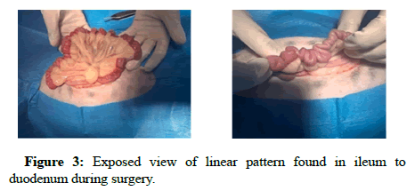
Clinical and surgical procedures performed in this case was exploratory laparotomy which is required to be performed to explore and to remove the liner foreign body. In pre-operative procedure medetomidine hydrochloride was used as pre-medication with ketamine combination for general anaesthesia which induces sedation. Medetomidine hydrochloride route of administration in cat’s only intramuscular injection with a dose of 0.05 ml-0.15 ml per kg body weight. While oh. Surprise. Ketamine drug’s route of administration can be intramuscular or intravenous injection with a dosage of 2.5 mg to 7.5 mg ketamine per kg body weight. Anaesthesia was maintained with 1.5% isoflurane. After the cat anesthetized and placed on a surgical table lying on her back, the hair was removed and the surgical field was cleaned and disinfected. A linear foreign body that is wrapped around the tongue should be identified and released before laparotomy, abdominal incision was made, the abdominal cavity was accessed through the ventral midline of the abdomen incision, the stomach was bloated due to barium and gases which were unable to pass. The linear FB may be localized by identifying intestinal complication. The intestine was in place and clear view of linear pattern found in ileum to duodenum (Figure 3), the knot caused obstruction in proximal part of ileum. After the location of the foreign body was determined and it can be pulled gently and gradually until the more distal point of fixation is reached.
Discussion
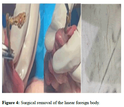
Multiple enterotoxins spaced along the intestine, are required to minimize excessive traction and avoid subsequent intestinal perforation and to remove the foreign body completely. However due to chronic ingestion of the threads, the threads were all over the small intestine which was removed (Figure 4).
The intestinal incision was sutured by cushing pattern, abdomen was sprayed several times with saline, complete closure of the abdomen, muscle and sub cutis by simple continuous suture pattern, the skin sutured by simple cross-mattress pattern. After that silver sulfadiazine spray was applied topically above the surgical wound which provides antimicrobial or antibacterial action and helps in healing process of the wound. In post-operative care, Metoclopramide Hydrochloride was prescribed which is commonly used to treat and prevent vomiting and to stimulate stomach and intestinal motility. It was administered by intramuscular injection route for cats with a dose of 0.2 mg per kg every six to eight hours. Meloxicam used to reduce inflammation and relief the pain. The route of administration is subcutaneous and the at a dose of 0.3 mg per kg body weight. Moreover, Sodium chloride 0.6% w/v intravenous infusion therapy was administered.
Follow up and outcome
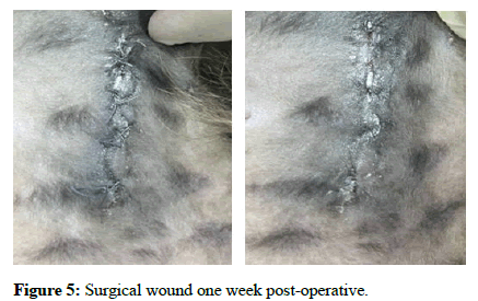
Prognosis will be good as long as the treatment proceeds well. The cat remained hospitalized for three days following the removal of this linear foreign body. During this time, the cat received intravenous fluids for hydration and pain medications to keep her comfortable. In the first 24 hours after surgery, the cat was fed small amounts of a bland and canned diet to help the gastrointestinal tract resume normal motility. The patient was observed closely to ensure that there is no sign of leakage at the intestinal incisions. Based on the response to this introduction of feed, the size of her meals was gradually increased until she is eating normally. We recommended that the owner should take proper precautions and prevent recurrence of this problem by removing any threads immediately and to keep eye on the cat. After the abdominal exploratory surgery, the animal was rested and advised to have a limited activity for at least 14 days to allow the surgical wound to heal. Elizabethan collar is necessary to prevent the cat from licking, opening or infecting the surgical wound to allow better healing and to prevent any further complications such as infection or break down of the wound. The surgery had positive impact and an uneventful recovery, the animal was presented to the clinic for follow up and to remove the suture (Figure 5). She was in good health status and had stopped vomiting, started to eat and recover her appetite gradually, the bowel movements were proper.
Conclusion
The linear foreign body is difficult to diagnose, strings are too small to visualize on a radiograph, examining under the tongue for a string is important but not always possible because sometimes even if the string is present, it is not always visible. The presence of plication on the radiograph or the paisley shaped gas patterns may help in the diagnosis. However, most of the times it is not definitive. Based on animal health status, exploratory laparotomy should be performed as treatment and diagnostic procedure. The final diagnosis of this case is linear foreign body based on history, clinical signs, physical examination and diagnostic tests and confirmed by exploratory laparotomy.
Acknowledgment
We would like to thank paws and claws veterinary clinic for their collaboration.
Ethical Considerations
The owner consent was taken prior to undertaking the treatment protocol following the standard procedures.
References
- Agudelo CFR, Filipejova Z, Frgelecova L, Sychra O (2018) An unusual foreign body in a cat: A case report. Vet Med 63:198-202.
- Bojrab MJ, Waldron DR, Toombs JP (2014) Current techniques in small animal surgery. 5th Edition. Teton NewMedia: USA. 1183.
- Evans KL, Smeak DD, Biller DS (1994) Gastrointestinal linear foreign bodies in 32 dogs: A retrospective evaluation and feline comparison. J Am Anim Hosp Assoc 30:445-450.
- Levitt L, Clark GR, Adams V (1993) Tracheal foreign body in a cat. Can Vet J 34:172-173.
[Google Scholar] [PubMed]
- Papazoglou LG, Patsikas MN, Rallis T (2003) Intestinal foreign bodies in dogs and cats. Compendium on continuing education for the practising veterinarian-North American edition 25:830-845.
- Saundra EW, Charles SF (1991) Partial gastrointestinal obstruction for one month due to a linear foreign body in a cat. Can Vet J 32:689-691.
[Google Scholar] [PubMed]
- Tenwolde AC, Johnson LR, Hunt GB, Vernau W, Zwingenberger AL (2010) The role of bronchoscopy in foreign body removal in dogs and cats: 37 cases (2000-2008). J Vet Intern Med 24:1063-1068.
[Crossref] [Google Scholar] [PubMed]
 Spanish
  Chinese
  Russian
  German
  French
  Japanese
  Portuguese
  Hindi
Hindi 