the guidewire inserted into the false lumen. (B) The IVUS catheter was advanced again over a second guidewire inserted into the true lumen. The first guidewire
inserted into the false lumen was observed (arrow). * - true lumen, # - false lumen.
 |
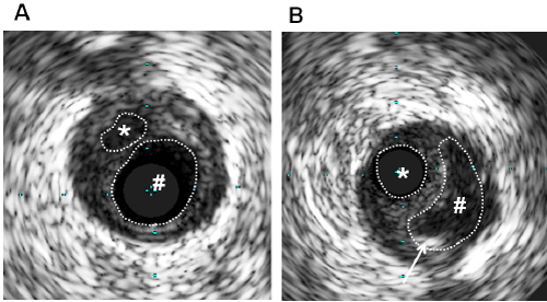
| Figure 2: Intravascular ultrasound (IVUS) imaging at the site of coronary artery dissection in the right coronary artery. (A) The IVUS catheter was advanced over the guidewire inserted into the false lumen. (B) The IVUS catheter was advanced again over a second guidewire inserted into the true lumen. The first guidewire inserted into the false lumen was observed (arrow). * - true lumen, # - false lumen. |