 |
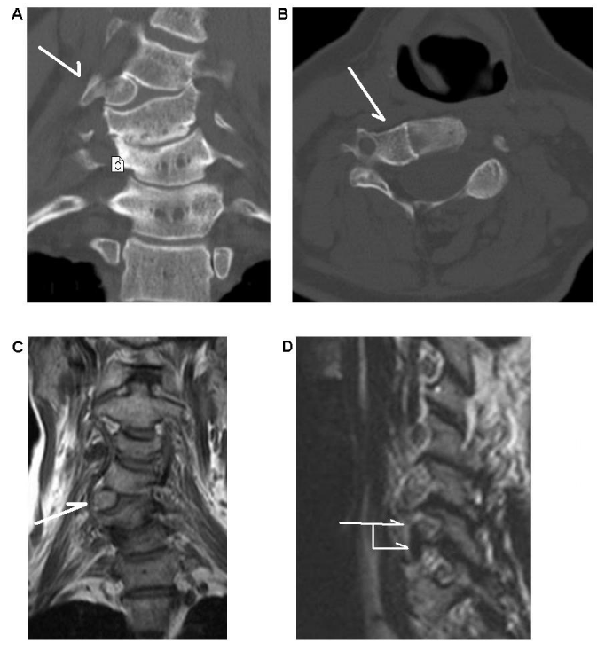
| Figure 1: A Cervical hemivertebra at the level of C5. A) The white arrow indicates the hemivertebra. At the adjacent level below, degenerative signs are seen on this CT image (coronal view). B) Transversal view of the hemivertebra. This bony anomaly has a completely developed vertebral canal and a neuroforamen. C) The MRI scan shows the hemivertebra and in D) it seems that above and below the hemivertebra nerve root foramina are present. |